

Comparison of the antiangiogenic activity of modified RGDRGD-endostatin to endostatin delivered by gene transfer in vivo rabbit neovascularization model
- PMID: 21850166
- PMCID: PMC3154123
Comparison of the antiangiogenic activity of modified RGDRGD-endostatin to endostatin delivered by gene transfer in vivo rabbit neovascularization model
Abstract
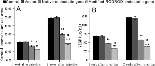
Purpose: Endostatin plays an important role in inhibiting corneal neovascularization (CNV). The aim of this study was to evaluate the antiangiogenic activities of lipid-mediated subconjunctival injection of the modified RGDRGD (arginine- glycin- aspartic- arginine- glycin- aspartic- endostatin gene in a rabbit model of neovascularization in vivo.
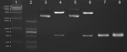
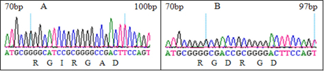
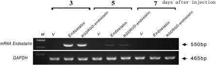
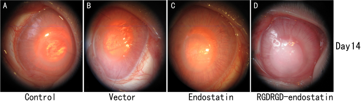
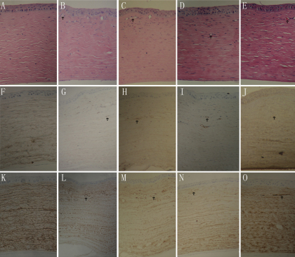
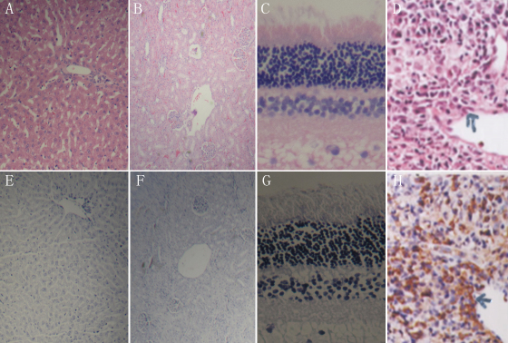
Methods: A modified human endostatin gene containing an RGDRGD motif was obtained by rapid site-directed mutagenesis. Forty New Zealand white rabbits underwent alkaline burn and developed CNV, which were randomly divided into four groups: an experimental control group, a PCI empty vector group, a PCI-endostatin group, and a PCI-RGDRGD-endostatin group. The vector, endostatin, and RGDRGD-endostatin groups received injections into the superior bulbar conjunctiva after the burn. An injection of 5 μg was given twice at 1-week intervals. Four eyes of two rabbits received neither treatment nor alkaline burn and served as absolute normal controls. The areas of CNV were monitored after 7 and 14 days. Corneas were examined by histology, and VEGF (vascular endothelial growth factor) and CD31 (platelet endothelial cell adhesion molecule-1) expression was detected by immunohistochemistry after 7 and 14 days. Retina, liver, and kidney were examined by histology, and CD38 expression in the inflammatory cells was detected by immunohistochemistry at 90 days.
Results: Subconjunctival injection of both native endostatin and modified RGDRGD-endostatin genes resulted in a significant suppression of CNV in vivo, with modified RGDRGD-endostatin being more effective than native endostatin. The mean concentration of VEGF in the PCI-RGDRGD-endostatin group significantly decreased compared to the means in the other groups. Upon histological examination, the endostatin-treated and RGDRGD-endostatin-treated eyes showed significantly less neovascular area and fewer vessels than the control and vector-injected groups. Retinal, hepatic, and renal tissue sections were normal, and there was no inflammatory cell infiltration observed.
Conclusions: Native and modified endostatin can significantly inhibit CNV by suppressing the expression of VEGF. However, modified endostatin with the RGDRGD motif is far more effective than the endostatin gene in antiangiogenic activity.
Figures
Similar articles
-
A C-terminal fragment BIGH3 protein with an RGDRGD motif inhibits corneal neovascularization in vitro and in vivo.Exp Eye Res. 2013 Jul;112:10-20. doi: 10.1016/j.exer.2013.03.014. Epub 2013 Apr 3. Exp Eye Res. 2013. PMID: 23562678
-
[Inhibition of corneal neovascularization by liposomes mediated plasmid encoding human endostatin].Zhonghua Yan Ke Za Zhi. 2005 Mar;41(3):260-4. Zhonghua Yan Ke Za Zhi. 2005. PMID: 15840371 Chinese.
-
Inhibition of mouse alkali burn induced-corneal neovascularization by recombinant adenovirus encoding human vasohibin-1.Mol Vis. 2010 Jul 26;16:1389-98. Mol Vis. 2010. PMID: 20680097 Free PMC article.
-
Recent drug therapies for corneal neovascularization.Chem Biol Drug Des. 2017 Nov;90(5):653-664. doi: 10.1111/cbdd.13018. Epub 2017 Jun 21. Chem Biol Drug Des. 2017. PMID: 28489275 Review.
-
Research progress on the correlation between corneal neovascularization and lymphangiogenesis (Review).Mol Med Rep. 2025 Feb;31(2):47. doi: 10.3892/mmr.2024.13412. Epub 2024 Dec 5. Mol Med Rep. 2025. PMID: 39635819 Free PMC article. Review.
Cited by
-
Doxycycline versus Curcumin for Inhibition of Matrix Metalloproteinase Expression and Activity Following Chemically Induced Inflammation in Corneal Cells.J Ophthalmic Vis Res. 2024 Sep 16;19(3):273-283. doi: 10.18502/jovr.v19i3.13689. eCollection 2024 Jul-Sep. J Ophthalmic Vis Res. 2024. PMID: 39359528 Free PMC article.
-
Anti-neovascular effect of chondrocyte-derived extracellular matrix on corneal alkaline burns in rabbits.Graefes Arch Clin Exp Ophthalmol. 2014 Jun;252(6):951-61. doi: 10.1007/s00417-014-2633-3. Epub 2014 May 1. Graefes Arch Clin Exp Ophthalmol. 2014. PMID: 24789464
-
Non-viral therapeutic approaches to ocular diseases: An overview and future directions.J Control Release. 2015 Dec 10;219:471-487. doi: 10.1016/j.jconrel.2015.10.007. Epub 2015 Oct 9. J Control Release. 2015. PMID: 26439665 Free PMC article. Review.
-
Management Strategies of Ocular Chemical Burns: Current Perspectives.Clin Ophthalmol. 2020 Sep 15;14:2687-2699. doi: 10.2147/OPTH.S235873. eCollection 2020. Clin Ophthalmol. 2020. PMID: 32982161 Free PMC article. Review.
-
AAV vector-meditated expression of HLA-G reduces injury-induced corneal vascularization, immune cell infiltration, and fibrosis.Sci Rep. 2017 Dec 19;7(1):17840. doi: 10.1038/s41598-017-18002-9. Sci Rep. 2017. PMID: 29259248 Free PMC article.
References
-
- Yoon KC, Ahn KY, Lee JH, Chun BJ, Park SW, Seo MS, Park YG, Kim KK. Lipid-mediated delivery of brain-specific angiogenesis inhibitor 1 gene reduces corneal neovascularization in an in vivo rabbit model. Gene Ther. 2005;12:617–24. - PubMed
-
- Chang JH, Gabison EE, Kato T, Azar DT. Corneal neovascularization. Curr Opin Ophthalmol. 2001;12:242–9. - PubMed
-
- Benelli U, Ross JR, Nardi M, Klintworth GK. Corneal neovascularization induced by xenografts or chemical cautery: Inhibition by cyclosporin A. Invest Ophthalmol Vis Sci. 1997;38:274–82. - PubMed
-
- Hayashi A, Popovich KS, Kim HC, de Juan E. Role of protein tyrosine phosphorylation in rat corneal neovascularization. Graefes Arch Clin Exp Ophthalmol. 1997;235:460–7. - PubMed
-
- Lee P, Wang CC, Adamis AP. Ocular neovascularization: an epidemiologic review. Surv Ophthalmol. 1998;43:245–69. - PubMed
Publication types
MeSH terms
Substances
LinkOut - more resources
Full Text Sources
Research Materials
Miscellaneous