. 2011:17:1946-56.
Epub 2011 Jul 19.

Intravitreal homocysteine-thiolactone injection leads to the degeneration of multiple retinal cells, including photoreceptors

Affiliations

Intravitreal homocysteine-thiolactone injection leads to the degeneration of multiple retinal cells, including photoreceptors

Han-Hsin Chang et al. Mol Vis. 2011.

Abstract

Purpose: Hyperhomocysteinemia is known to cause degeneration of retinal ganglion cells, but its influence on photoreceptors remains largely unknown. In particular, the role of homocysteine-thiolactone (Hcy-T)--the physiologic metabolite of homocysteine that has been proven to be more cytotoxic than homocysteine itself--as a factor that causes retinopathy, has not been defined. This study aimed to investigate the toxic effects of excessive Hcy-T in a mouse model.

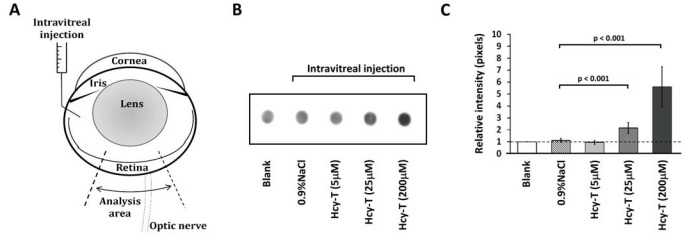
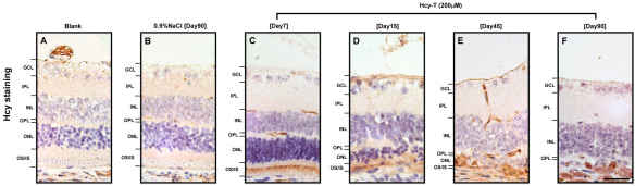
Methods: A total of 60 six-week-old female ICR mice were used in this study. The mice were divided into 3 experimental groups and 2 control groups. The mice in the experimental groups were subjected to intravitreal injections of Hcy-T to reach final estimated intravitreal concentrations at 5, 25, and 200 μM, respectively. Mice without injection (blank) and with 0.9 NaCl injections (sham injection) were used as controls. The mice with 200 μM Hcy-T were sacrificed at days 7, 15, 45, and 90 after injection and the mice with 5 or 25 μM Hcy-T were sacrificed at day 90, with the controls sacrificed at day 15 or 90 for comparison. Semi-quantitative dot-blot analysis was performed for confirmation of retinal homocysteinylation. The mouse retinas were evaluated microscopically, with the thickness of total and specific retinal layers determined. Immunohistochemical analysis was performed and the labeled cells were quantified to determine the effects of excessive Hcy-T on specific retinal cells.

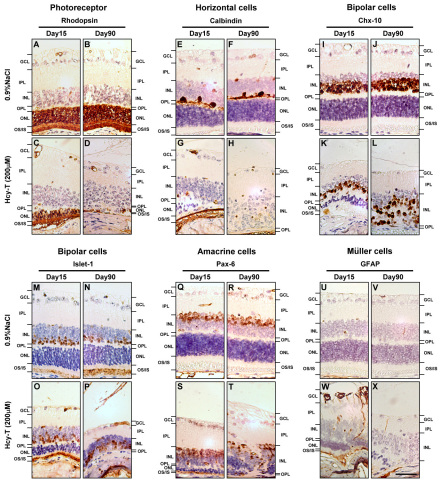
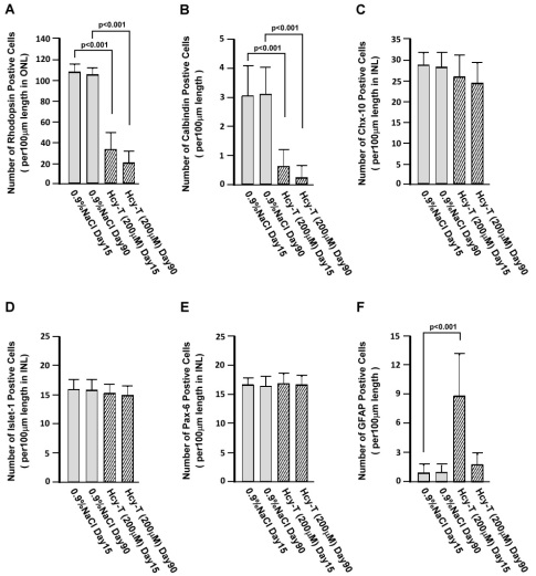
Results: Dose-dependent retinal homocysteinylation after Hcy-T injection was confirmed. The homocysteinylation was localized in the outer and inner segments of photoreceptors and the ganglion cell layer (GCL). Retinal cell degenerations were found in the GCL, inner nuclear layer, and outer nuclear layer at day 90 after 200 µM Hcy-T injection. Significant thickness reduction was found in the total retina, outer nuclear layer, and the outer and inner segment layers. A trend of thickness reduction was also found in the GCL and inner nuclear layer, although this was not statistically significant. The rhodopsin⁺ photoreceptors and the calbindin⁺ horizontal cells were significantly reduced at day 15, and were nearly ablated at day 90 after 200 μM Hcy-T injection (p<0.001 for both day 15 and day 90), which was not seen in the sham injection controls. The Chx-10⁺ or the Islet-1⁺ bipolar cells and the Pax-6⁺ amacrine cells were severely misarranged at day 90, but no significant reduction was found for both cell types. The GFAP⁺ Müller cells were activated at day 15, but were not significantly increased at day 90 after the injection.

Conclusions: Excessive retinal homocysteinylation by Hcy-T, a condition of hyperhomocysteinemia, could lead to degeneration of photoreceptors, which might lead to retinopathies associated with severe hyperhomocysteinemia or diabetes mellitus.


Figures

Figure 1
Figure 1
The preparation of intravitreal homocysteine-thiolactone (Hcy-T) injection is illustrated in A and retinal homocystinylation as confirmed by a semi-quantitative dot-blot analysis is shown in B and C. A: In each retinal section, the thickness measurement was made at two sites, located approximately 200 to 300 μm apart, on either side of the optic nerve. B: Dose-dependent retinal homocystinylation was observed on day 90 after injection. C: Quantification of relative intensity (pixels) on dot-blot membranes using an arbitrary unit of retinal homocystinylation as compared to that of the blank control. The sample size was 5 in all groups. The error bars indicate standard error of the means (SEMs).
Figure 2
Figure 2
The effects of an intravitreal homocysteine-thiolactone (Hcy-T) injection on mouse retinas at day 90 after preparation. All retinas were prepared with hematoxylin-eosin staining for histological assessment and for measurement of the thickness of specific layers. A: Blank control without any injection. B: 0.9% NaCl sham injection control. C: Final intravitreal Hcy-T at 5 μM. D: Final intravitreal Hcy-T at 25 μM. E: Final intravitreal Hcy-T at 200 μM. F: Total retinal thickness. G: Ganglion cell layer (GCL) thickness. H: Inner nuclear layer (INL) thickness. I: Outer nuclear layer (ONL) thickness. J: Outer and inner segments of photoreceptors (OS/IS) thickness. The sample size for total retinal thickness was 5 in all groups. The error bars indicate standard error of the means (SEMs). Scale bar: 25 μm.
Figure 3
Figure 3
The immunohistochemical localization of retinal homocystinylation sites at days 7, 15, 45, 90 after a homocysteine-thiolactone (Hcy-T) injection to reach a final intravitreal Hcy-T concentration at 200 μM. A: Blank control without any injection. B: 0.9% NaCl sham injection control at day 90. C: Day 7 after injection. D: Day 15 after injection. E: Day 45 after injection. F: Day 90 after injection. Scale bar: 40 μm.
Figure 4
Figure 4
The cytotoxic effects of homocysteine-thiolactone (Hcy-T) on specific retinal cells as reflected by alterations of A-D: rhodopsin positive photoreceptors, E-H: calbindin positive horizontal cells, I-L: Chx- 10 positive bipolar cells, M-P: Islet-1 positive bipolar cells, Q-T: Pax-6 positive amacrine cells, and U-X: glial fibrillary acidic protein (GFAP) positive Müller cells at day 15 and day 90 following the intravitreal injections. Abbreviations: GCLrepresents ganglion cell layer; INL represents inner nuclear layer; IPL represents inner plexiform layer; ONL represents outer nuclear layer; OPL represents outer plexiform layer; OS/IS represents outer and inner segments of photoreceptors. Scale bar: 45 μm.
Figure 5
Figure 5
Quantification of specific retinal cells per 100 μm length according to their specific markers. The markers were used to detect at day 90 following the intravitreal injections. A: Rhodopsin positive cells per 100 μm length in outer nuclear layer (ONL). B: Calbindin positive cells per 100 μm length in the retina. C: Chx-10 positive cells per 100 μm length in inner nuclear layer (INL). D: Islet-1 positive cells per 100 μm length in INL. E: Pax-6 positive cells per 100 μm length in INL. F: Glial fibrillary acidic protein (GFAP) positive cells per 100 μm length in the retina. The sample size for all markers was 5 in all groups. The error bars indicate standard error of the means (SEMs).

References

    1. Perla-Kaján J, Twardowski T, Jakubowski H. Mechanisms of homocysteine toxicity in humans. Amino Acids. 2007;32:561–72. - PubMed
    1. Karolczak K, Olas B. Mechanism of action of homocysteine and its thiolactone in hemostasis system. Physiol Res. 2009;58:623–33. - PubMed
    1. Dionisio N, Jardin I, Salido GM, Rosado JA. Homocysteine, intracellular signaling and thrombotic disorders. Curr Med Chem. 2010;17:3109–19. - PubMed
    1. Ghorbanihaghjo A, Javadzadeh A, Argani H, Nezami N, Rashtchizadeh N, Rafeey M, Rohbaninoubar M, Rahimi-Ardabili B. Lipoprotein(a), homocysteine, and retinal arteriosclerosis. Mol Vis. 2008;14:1692–7. - PMC - PubMed
    1. Micheal S, Qamar R, Akhtar F, Khan MI, Khan WA, Ahmed A. MTHFR gene C677T and A1298C polymorphisms and homocysteine levels in primary open angle and primary closed angle glaucoma. Mol Vis. 2009;15:2268–78. - PMC - PubMed

Publication types

MeSH terms

LinkOut - more resources