

Critical role of SDF-1α-induced progenitor cell recruitment and macrophage VEGF production in the experimental corneal neovascularization
- PMID: 21850188
- PMCID: PMC3156784
Critical role of SDF-1α-induced progenitor cell recruitment and macrophage VEGF production in the experimental corneal neovascularization
Abstract
Purpose: To address the roles of the stromal derived factor-1 (SDF-1) α in the course of experimental corneal neovascularization (CNV).
Methods: CNV was induced by alkali injury and compared in SDF-1α- or vehicle-treated mice two weeks after injury. Angiogenic factor expression in the early phase after injury was quantified by reverse transcription polymerase chain reaction (RT-PCR). Progenitor cell, macrophage, and monocyte intracorneal accumulation in the early phase after injury was evaluated by flow cytometric analysis.
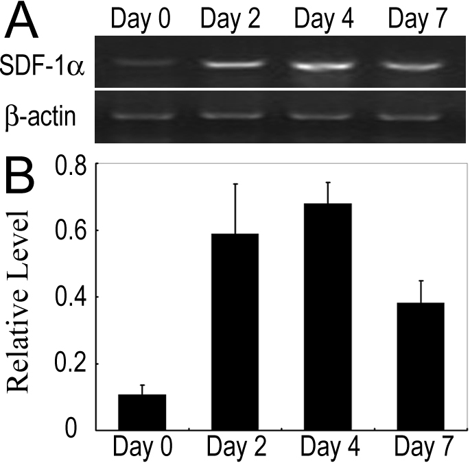
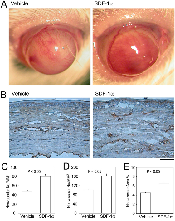
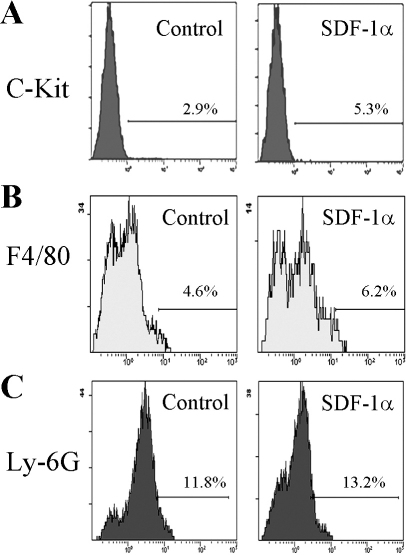
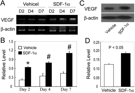
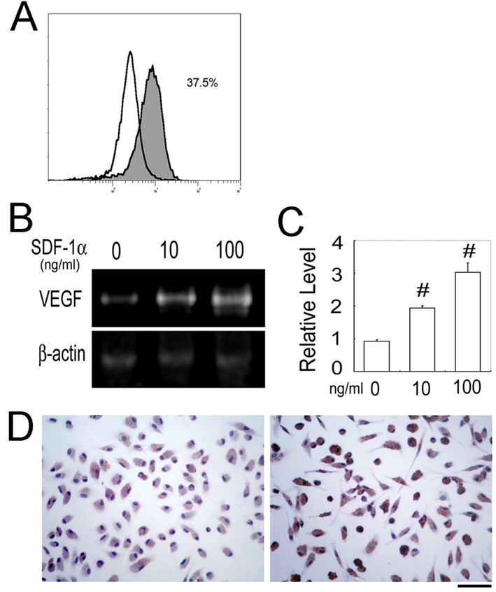
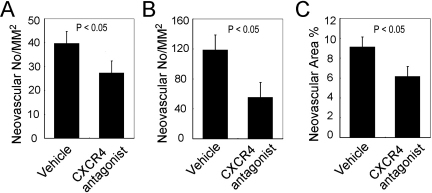
Results: The mRNA expression of SDF-1α was augmented, together with infiltration of c-kit-positive progenitor cells in the corneas after the alkali injury. Compared with vehicle-treated mice, SDF-1α-treated mice exhibited enhanced CNV two weeks after injury, as evidenced by enlarged cluster of differentiation 31 (CD31)-positive areas. Concomitantly, the intracorneal infiltration of c-kit-positive progenitor cells but not F4/80+ macrophages or Ly-6G+ monocytes was significantly enhanced in SDF-1α-treated mice compared to vehicle-treated mice. SDF-1α enhanced vascular endothelial growth factor (VEGF) expression by murine peritoneal macrophages. Enhancement in intraocular VEGF expression was greater in SDF-1α-treated mice than in control mice after injury. Moreover, local administration of C-X-C chemokine receptor type 4 (CXCR4) antagonist after alkali injury reduced alkali-induced CNV.
Conclusions: SDF-1α-treated mice exhibited enhanced alkali-induced CNV through enhanced intracorneal progenitor cell infiltration and increased VEGF expression by macrophages.
Figures
Similar articles
-
Critical role of TNF-α-induced macrophage VEGF and iNOS production in the experimental corneal neovascularization.Invest Ophthalmol Vis Sci. 2012 Jun 14;53(7):3516-26. doi: 10.1167/iovs.10-5548. Invest Ophthalmol Vis Sci. 2012. PMID: 22570350
-
Essential contribution of CCL3 to alkali-induced corneal neovascularization by regulating vascular endothelial growth factor production by macrophages.Mol Vis. 2008 Sep 5;14:1614-22. Mol Vis. 2008. PMID: 18776949 Free PMC article.
-
Enhanced experimental corneal neovascularization along with aberrant angiogenic factor expression in the absence of IL-1 receptor antagonist.Invest Ophthalmol Vis Sci. 2009 Oct;50(10):4761-8. doi: 10.1167/iovs.08-2732. Epub 2009 May 20. Invest Ophthalmol Vis Sci. 2009. PMID: 19458323
-
Genetically manipulated progenitor/stem cells restore function to the infarcted heart via the SDF-1α/CXCR4 signaling pathway.Prog Mol Biol Transl Sci. 2012;111:265-84. doi: 10.1016/B978-0-12-398459-3.00012-5. Prog Mol Biol Transl Sci. 2012. PMID: 22917235 Review.
-
Concise review: the potential of stromal cell-derived factor 1 and its receptors to promote stem cell functions in spinal cord repair.Stem Cells Transl Med. 2012 Oct;1(10):732-9. doi: 10.5966/sctm.2012-0068. Epub 2012 Oct 10. Stem Cells Transl Med. 2012. PMID: 23197665 Free PMC article. Review.
Cited by
-
Inhibition of angiogenesis, fibrosis and thrombosis by tetramethylpyrazine: mechanisms contributing to the SDF-1/CXCR4 axis.PLoS One. 2014 Feb 5;9(2):e88176. doi: 10.1371/journal.pone.0088176. eCollection 2014. PLoS One. 2014. PMID: 24505417 Free PMC article.
-
SDF‑1α/CXCR4 signaling promotes capillary tube formation of human retinal vascular endothelial cells by activating ERK1/2 and PI3K pathways in vitro.Mol Med Rep. 2022 Oct;26(4):305. doi: 10.3892/mmr.2022.12821. Epub 2022 Aug 10. Mol Med Rep. 2022. PMID: 35946444 Free PMC article.
-
Pathogenesis of Alkali Injury-Induced Limbal Stem Cell Deficiency: A Literature Survey of Animal Models.Cells. 2023 May 1;12(9):1294. doi: 10.3390/cells12091294. Cells. 2023. PMID: 37174694 Free PMC article. Review.
-
Anti-apoptosis effects of vascular endothelial cadherin in experimental corneal neovascularization.Int J Ophthalmol. 2015 Dec 18;8(6):1083-8. doi: 10.3980/j.issn.2222-3959.2015.06.01. eCollection 2015. Int J Ophthalmol. 2015. PMID: 26682152 Free PMC article.
-
Effects of CXCR4 gene silencing by lentivirus shRNA on proliferation of the EC9706 human esophageal carcinoma cell line.Tumour Biol. 2013 Oct;34(5):2951-9. doi: 10.1007/s13277-013-0858-0. Epub 2013 Jun 7. Tumour Biol. 2013. PMID: 23744460
References
-
- Ambati BK, Nozaki M, Singh N, Takeda A, Jani PD, Suthar T, Albuquerque RJC, Richter E, Sakurai E, Newcomb MT, Kleinman ME, Caldwell RB, Lin Q, Ogura Y, Orecchia A, Samuelson DA, Agnew DW, St. Leger J, Green WR, Mahasreshti PJ, Curiel DT, Kwan D, Marsh H, Ikeda S, Leiper LJ, Collinson JM, Bogdanovich S, Khurana TS, Shibuya M, Baldwin ME, Ferrara N, Gerber H-P, DeFalco S, Witta J, Baffi JZ, Raisler BJ, Ambati J. Corneal avascularity is due to soluble VEGF receptor-1. Nature. 2006;443:993–7. - PMC - PubMed
-
- Cursiefen C, Masli S, Ng TF, Dana MR, Bornstein P, Lawler J, Streilein JW. Roles of thrombospondin-1 and −2 in regulating corneal and iris angiogenesis. Invest Ophthalmol Vis Sci. 2004;45:1117–24. - PubMed
-
- Gao G, Ma J. Tipping the balance for angiogenic disorders. Drug Discov Today. 2002;7:171–2. - PubMed
-
- Zhang SX, Ma JX. Ocular neovascularization: Implication of endogenous angiogenic inhibitors and potential therapy. Prog Retin Eye Res. 2007;26:1–37. - PubMed
-
- Lu P, Li L, Mukaida N, Zhang X. Alkali-induced corneal neovascularization is independent of CXCR2-mediated neutrophil infiltration. Cornea. 2007;26:199–206. - PubMed
Publication types
MeSH terms
Substances
LinkOut - more resources
Full Text Sources