

A voxel-based morphometric analysis of cerebral gray matter in subcortical ischemic vascular dementia patients and normal aged controls
- PMID: 21850200
- PMCID: PMC3156997
- DOI: 10.7150/ijms.8.482
A voxel-based morphometric analysis of cerebral gray matter in subcortical ischemic vascular dementia patients and normal aged controls
Abstract
Background and purpose: The present study was designed to detect the abnormalities of the cerebral grey-matter density in subcortical ischemic vascular dementia patients by FSL-VBM method to promote the early diagnosis of it.
Methods: Nine subcortical ischemic vascular dementia patients and nine age-matched normal controls underwent MRI brain structure scanning that was performed on a SIEMENS AVANTO 1.5 Tesla scanner and standard T1-weighted high-resolution anatomic scans of MPRAGE sequence were obtained. The 3-demensional MPRAGE images were processed with FSL-VBM package and the cerebral gray matter density was compared between the subcortical ischemic vascular dementia patients and normal controls.
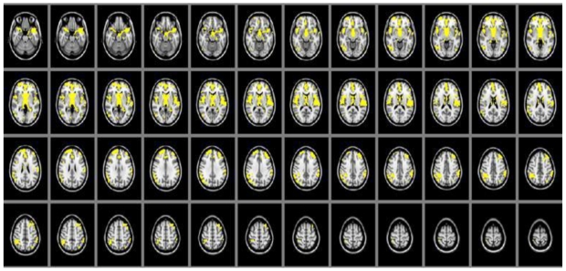
Results: Compared with the normal control group, the cerebral gray matter density of subcortical ischemic vascular dementia patients was found significantly decreasing, including brain regions of thalamus, parietal lobe, frontal lobe and temporal lobe (P<0.05).
Conclusions: The cerebral gray matter density alterations have closed correlation with cognitive dysfunction in subcortical ischemic vascular dementia patient and can be detected by MRI. MRI has some potential value in the diagnosis of them.
Keywords: MRI; cerebral grey-matter density; subcortical ischemic vascular dementia.
Conflict of interest statement
Conflict of Interest: The authors have declared that no conflict of interest exists.
Figures
Similar articles
-
The pattern of brain gray matter impairments in patients with subcortical vascular dementia.J Neurol Sci. 2014 Jun 15;341(1-2):110-8. doi: 10.1016/j.jns.2014.04.017. Epub 2014 Apr 24. J Neurol Sci. 2014. PMID: 24798224
-
Changes of brain structure in Parkinson's disease patients with mild cognitive impairment analyzed via VBM technology.Neurosci Lett. 2017 Sep 29;658:121-132. doi: 10.1016/j.neulet.2017.08.028. Epub 2017 Aug 18. Neurosci Lett. 2017. PMID: 28823894
-
Cerebral gray matter volume reduction in subcortical vascular mild cognitive impairment patients and subcortical vascular dementia patients, and its relation with cognitive deficits.Brain Behav. 2017 Jun 23;7(8):e00745. doi: 10.1002/brb3.745. eCollection 2017 Aug. Brain Behav. 2017. PMID: 28828207 Free PMC article.
-
Study of gray matter atrophy pattern with subcortical ischemic vascular disease-vascular cognitive impairment no dementia based on structural magnetic resonance imaging.Front Aging Neurosci. 2023 Feb 6;15:1051177. doi: 10.3389/fnagi.2023.1051177. eCollection 2023. Front Aging Neurosci. 2023. PMID: 36815175 Free PMC article.
-
Cortical and Subcortical Gray Matter Volume in Youths With Conduct Problems: A Meta-analysis.JAMA Psychiatry. 2016 Jan;73(1):64-72. doi: 10.1001/jamapsychiatry.2015.2423. JAMA Psychiatry. 2016. PMID: 26650724 Review.
Cited by
-
No increased risk of dementia in patients receiving androgen deprivation therapy for prostate cancer: a 5-year follow-up study.Asian J Androl. 2017 Jul-Aug;19(4):414-417. doi: 10.4103/1008-682X.179528. Asian J Androl. 2017. PMID: 27232853 Free PMC article.
-
Alterations of the cerebral cortex in sporadic small vessel disease: A systematic review of in vivo MRI data.J Cereb Blood Flow Metab. 2016 Apr;36(4):681-95. doi: 10.1177/0271678X15625352. Epub 2016 Jan 19. J Cereb Blood Flow Metab. 2016. PMID: 26787108 Free PMC article.
-
Leukoaraiosis and Gray Matter Volume Alteration in Older Adults: The PROOF Study.Front Neurosci. 2022 Jan 13;15:747569. doi: 10.3389/fnins.2021.747569. eCollection 2021. Front Neurosci. 2022. PMID: 35095388 Free PMC article.
-
A panel of clinical and neuropathological features of cerebrovascular disease through the novel neuroimaging methods.Dement Neuropsychol. 2017 Oct-Dec;11(4):343-355. doi: 10.1590/1980-57642016dn11-040003. Dement Neuropsychol. 2017. PMID: 29354214 Free PMC article.
-
Neuroimaging biomarkers of neurodegenerative diseases and dementia.Semin Neurol. 2013 Sep;33(4):386-416. doi: 10.1055/s-0033-1359312. Epub 2013 Nov 14. Semin Neurol. 2013. PMID: 24234359 Free PMC article. Review.
References
-
- Jellinger KA. The enigma of vascular cognitive disorder and vascular dementia. Acta Neuropathol. 2007;113:349–88. - PubMed
-
- Pohjasvaara T, Mantyla R, Ylikoski R. et al. Comparison of different clinical criteria (DSM-III, ADDTC, ICD-10, NINDS-AIREN, DSM-IV) for the diagnosis of vascular dementia. National Institute of Neurological Disorders and Stroke-Association Internationale pour la Recherche et l'Enseignement en Neurosciences. Stroke. 2000;31:2952–7. - PubMed
-
- Ross GW, Petrovitch H, White LR. et al. Characterization of risk factors for vascular dementia: The Honolulu Asia Aging Study. Neurology. 1999;53:337–43. - PubMed
-
- Loeb C, Gandolfo C, Croce R. et al. Dementia associated with lacunar infarction. Stroke. 1992;23:1225–9. - PubMed
Publication types
MeSH terms
LinkOut - more resources
Full Text Sources
