

doi: 10.1371/journal.pone.0022394.
Epub 2011 Aug 5.
Simple, time-saving dye staining of proteins for sodium dodecyl sulfate-polyacrylamide gel electrophoresis using Coomassie blue
Affiliations
- PMID: 21850222
- PMCID: PMC3151240
- DOI: 10.1371/journal.pone.0022394
Item in Clipboard
Simple, time-saving dye staining of proteins for sodium dodecyl sulfate-polyacrylamide gel electrophoresis using Coomassie blue
PLoS One.
2011.
Abstract
A fixation-free and fast protein-staining method for sodium dodecyl sulfate-polyacrylamide gel electrophoresis using Coomassie blue is described. The protocol comprises staining and quick washing steps, which can be completed in 0.5 h. It has a sensitivity of 10 ng, comparable with that of conventional Coomassie Brilliant Blue G staining with phosphoric acid in the staining solution. In addition, the dye stain does not contain any amount of acid and methanol, such as phosphoric acid. Considering the speed, simplicity, and low cost, the dye stain may be of more practical value than other dye-based protein stains in routine proteomic research.
Conflict of interest statement
Figures
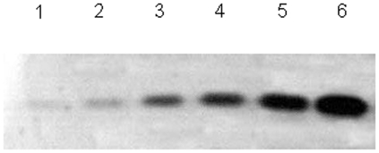
Different concentrations of BSA were separated by SDS-PAGE and stained with water-soluble CBBR at boiling temperature for 60 s. Lanes 1–6 represent 10 ng, 100 ng, 500 ng, 1 µg, 5 µg, and 10 µg of BSA.
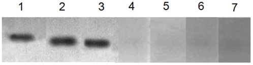
BSA (500 ng) was separated by SDS-PAGE and stained with different acid-soluble CBBR at boiling temperature for 60 s. Added to the water-soluble CBBR staining solution were 0.3% HCl (Lane 1), 0.3% phosphoric acid (Lane 2), and 0.3% acetic acid (Lane 4), respectively.
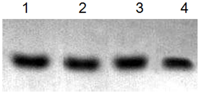
BSA (500 ng) was separated by SDS-PAGE and stained with water-soluble CBBR solution at boiling temperature for 30 s (Lane 1), 60 s (Lane 2), and 120 s (Lane 3) and at room temperature for 1 h (Lane 4), 6 h (Lane 5), 14 h (Lane 6), and 24 h (Lane 7).
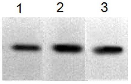
BSA (500 ng) was separated by SDS-PAGE, and the separated gels were washed with different types of water: running water (Lane 1), distilled water (Lane 2), and double-distilled water (Lane 3) and stained with water-soluble CBBR solution at boiling temperature for 60 s.
Similar articles
-
A Simple, Time-Saving Dye Staining of Proteins in Sodium Dodecyl Sulfate-Polyacrylamide Gel Using Coomassie Blue.Methods Mol Biol. 2018;1853:31-35. doi: 10.1007/978-1-4939-8745-0_5. Methods Mol Biol. 2018. PMID: 30097927
-
Counterion dye staining of proteins in one- and two-dimensional sodium dodecyl sulfate-polyacrylamide gel electrophoresis and tryptic gel digestion of stained protein for mass spectrometry.Methods Mol Biol. 2012;869:497-509. doi: 10.1007/978-1-61779-821-4_44. Methods Mol Biol. 2012. PMID: 22585515
-
Counterion Dye Staining of Proteins in One- and Two-Dimensional Sodium Dodecyl Sulfate-Polyacrylamide Gel Electrophoresis and Tryptic Gel Digestion of Stained Protein for Mass Spectrometry.Methods Mol Biol. 2018;1853:53-64. doi: 10.1007/978-1-4939-8745-0_8. Methods Mol Biol. 2018. PMID: 30097930
-
[Protein determination by binding with the dye Coomassie brilliant blue G-250].Biokhimiia. 1994 Jun;59(6):763-77. Biokhimiia. 1994. PMID: 7521220 Review. Russian.
-
A brief review of other notable protein detection methods on acrylamide gels.Methods Mol Biol. 2012;869:617-20. doi: 10.1007/978-1-61779-821-4_56. Methods Mol Biol. 2012. PMID: 22585527 Free PMC article. Review.
Cited by
-
Comparative proteomic analysis of the stolon cold stress response between the C4 perennial grass species Zoysia japonica and Zoysia metrella.PLoS One. 2013 Sep 26;8(9):e75705. doi: 10.1371/journal.pone.0075705. eCollection 2013. PLoS One. 2013. PMID: 24086619 Free PMC article.
-
A general fluorescent light-up probe for staining and quantifying protein.R Soc Open Sci. 2019 Aug 28;6(8):190580. doi: 10.1098/rsos.190580. eCollection 2019 Aug. R Soc Open Sci. 2019. PMID: 31598246 Free PMC article.
-
Overexpression of suppressor of IKBKE 1 is associated with vincristine resistance in colon cancer cells.Biomed Rep. 2016 Nov;5(5):585-588. doi: 10.3892/br.2016.759. Epub 2016 Sep 21. Biomed Rep. 2016. PMID: 27882221 Free PMC article.
-
Effects of Social Defeat Stress on Microtubule Regulating Proteins and Tubulin Polymerization.Clin Psychopharmacol Neurosci. 2024 Feb 29;22(1):129-138. doi: 10.9758/cpn.23.1077. Epub 2023 Aug 10. Clin Psychopharmacol Neurosci. 2024. PMID: 38247419 Free PMC article.
-
BRCA1 expression serves a role in vincristine resistance in colon cancer cells.Oncol Lett. 2017 Jul;14(1):345-348. doi: 10.3892/ol.2017.6149. Epub 2017 May 10. Oncol Lett. 2017. PMID: 28693174 Free PMC article.
References
-
- Walker J, editor. 2002. pp. 61–67. (2002) The Protein Protocols Handbook (2nd Ed.) Humana Press Totowa.
-
- Shofuda K, Nagashima Y, Kawahara K, Yasumitsu H, Miki K, et al. Elevated expression of membrane-type 1 and 3 matrix metalloproteinases in rat vascular smooth muscle cells activated by arterial injury. Lab Invest. 1998;78:915–923. - PubMed
-
- Kawsar S, Fujii Y, Matsumoto R, Ichikawa T, Tateno H, et al. Isolation, purification, characterization and glycan-binding profile of a d-galactoside specific lectin from the marine sponge, Halichondria okadai. Comp Biochem Physiol B Biochem Mol Biol. 2008;150:349–357. - PubMed
-
- Kawsar S, Takeuchi T, Kasai K, Fujii Y, Matsumoto R, et al. Glycan-binding profile of a D-galactose binding lectin purified from the annelid, Perinereis nuntia ver. vallata. Comp Biochem Physiol B Biochem Mol Biol. 2009;152:382–389. - PubMed
-
- Yasumitsu H, Ozeki Y, Kawasar SM, Fujii Y, Sakagami M, et al. RAMA stain: A fast, sensitive and less protein-modifying CBB R250 stain. Electrophoresis. 2010;31:1913–1917. - PubMed
Publication types
MeSH terms
Substances
LinkOut - more resources
Full Text Sources
