. 2011;6(8):e22523.
doi: 10.1371/journal.pone.0022523. Epub 2011 Aug 5.

High-throughput identification of potential minor histocompatibility antigens by MHC tetramer-based screening: feasibility and limitations

Affiliations

High-throughput identification of potential minor histocompatibility antigens by MHC tetramer-based screening: feasibility and limitations

Pleun Hombrink et al. PLoS One. 2011.

Abstract

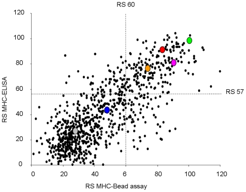
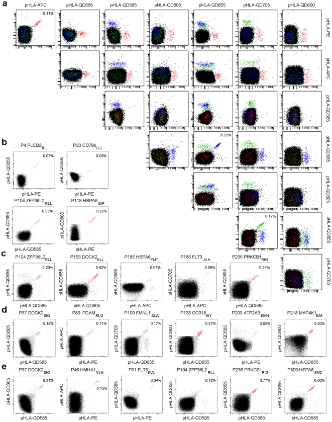
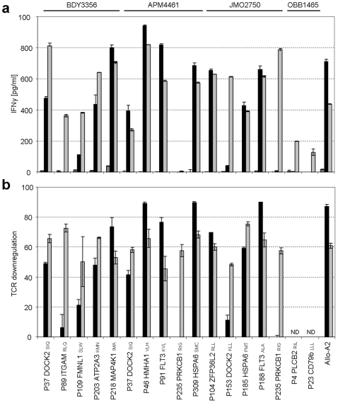
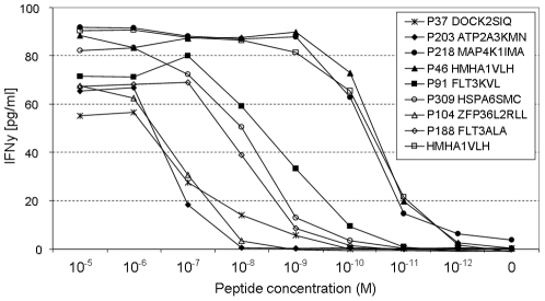
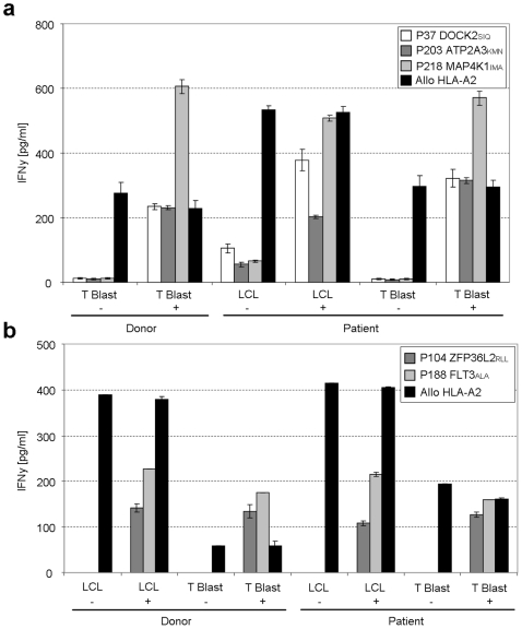
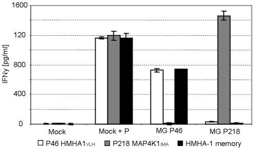
T-cell recognition of minor histocompatibility antigens (MiHA) plays an important role in the graft-versus-tumor (GVT) effect of allogeneic stem cell transplantation (allo-SCT). However, the number of MiHA identified to date remains limited, making clinical application of MiHA reactive T-cell infusion difficult. This study represents the first attempt of genome-wide prediction of MiHA, coupled to the isolation of T-cell populations that react with these antigens. In this unbiased high-throughput MiHA screen, both the possibilities and pitfalls of this approach were investigated. First, 973 polymorphic peptides expressed by hematopoietic stem cells were predicted and screened for HLA-A2 binding. Subsequently a set of 333 high affinity HLA-A2 ligands was identified and post transplantation samples from allo-SCT patients were screened for T-cell reactivity by a combination of pMHC-tetramer-based enrichment and multi-color flow cytometry. Using this approach, 71 peptide-reactive T-cell populations were generated. The isolation of a T-cell line specifically recognizing target cells expressing the MAP4K1(IMA) antigen demonstrates that identification of MiHA through this approach is in principle feasible. However, with the exception of the known MiHA HMHA1, none of the other T-cell populations that were generated demonstrated recognition of endogenously MiHA expressing target cells, even though recognition of peptide-loaded targets was often apparent. Collectively these results demonstrate the technical feasibility of high-throughput analysis of antigen-specific T-cell responses in small patient samples. However, the high-sensitivity of this approach requires the use of potential epitope sets that are not solely based on MHC binding, to prevent the frequent detection of T-cell responses that lack biological relevance.


Conflict of interest statement

Competing Interests: The authors have declared that no competing interests exist.

Figures

Figure 1
Figure 1. HLA-A2 affinity of predicted peptides measured in parallel by two different assays.
HLA-A2 affinity of 973 predicted MiHA peptides was measured in parallel by two different binding assays. Each dot (black) represents a pMHC complex rescued by a tested peptide after UV induced cleavage of a conditional ligand. On the y-axis rescue score (RS) are shown for MHC-ELISA assay. On the x-axis RS are shown for MHC-bead assay. RS are normalized to the HLA-A2 high affinity CMV-pp65NLV peptide and CMV-pp65NLV peptide RS set to 100 for both assays. Selection threshold: RS≥57 (MHC-ELISA) and RS≥60 (MHC-bead assay). High affinity peptide controls: CMV-NLVPMVATV (green), FLU-GILGFVFTL (pink), EBV-GLCTLVAML (orange) and HA1-VLHDDLLEA (red). Low affinity peptide control: MART1-AAGIGILTV (blue) and negative control A3-gp100-LIYRRRLMK (grey).
Figure 2
Figure 2. Detection of potential MiHA specific T-cell populations by pMHC tetramer staining.
These FACS analyses show the detection of MiHA specific T-cell populations through dual-encoding after pMHC tetramer pull down and in vitro expansion. Shown are total CD8+ T-cells. All dot plots are shown with bi-exponential axes and display fluorescence intensity for the indicated fluorochromes at the top and right of the plot matrix. Non-pMHC tetramer specific CD8+ T-cells are indicated black. Dot plots of pMHC tetramer positive T-cell populations are shown by staining one expanded cell culture with 16 separate panels of up to 25 different dual-color pMHC tetramers. (a) Representative example of pMHC multimer screen panel 5, patient BDY3356. Detection of three dual-labeled potential MiHA specific T-cell populations: P89 ITGAMRLQ (red), P104 ZFP36L2RLL (blue) and P109 FMNL1SLW (green). Frequencies indicate MiHA specific T-cells of total CD8+ cells. A selection of 21 potential MiHA specific T-cell populations was made with the highest clinical potential. Selected T-cell populations were derived from allo-SCT patient: OBB1465 (b), JMO2750 (c), BDY3356 (d) and APM4461 (e). Dot plots shown are representative for all detected dual-positive CD8+ T-cell populations (red).
Figure 3
Figure 3. Peptide stimulation leads to IFN γ production and TCR downregulation for 10 out of 17 pMHC tetramer positive cell lines.
Isolated pMHC tetramer positive cell lines were stimulated with peptide-loaded HLA-A2+ T2 target cells for 18 hours. Data is shown for 17 cell lines that were successfully generated by flowcytometry based cell sorting. Tested cell lines were derived from four different allo-SCT patients as indicated at the top of the graph. As a control an alloreactive CTL clone specific for a HLA-A2 epitope was used (Allo-A2). (a) Antigen specificity and functionality was analyzed by cytokine secretion in a standard IFN γ ELISA. Cell lines were stimulated with non-peptide loaded T2 cells (dark grey), [1 ug/ml] peptide-loaded T2 cells (black) and αCD3/CD28 stimulation beads (light grey). Data are presented as cytokine concentration. (b) Antigen specificity and functionality was analyzed by TCR internalization upon peptide stimulation. Cell lines shown were stimulated with [1 ug/ml] peptide-loaded T2 cells (black) and αCD3/CD28 stimulation beads (light grey). TCR downregulation was normalized to stimulation with non-peptide loaded T2 cell controls. Experiments were performed in duplicate, data are mean ± SD.
Figure 4
Figure 4. Analysis of peptide affinity of pMHC tetramer positive cell lines MHC tetramer positive T-cell lines demonstrated a wide range of peptide sensitivity.
HLA A2+ T2 cells were pulsed with specific MiHA peptide. Peptide concentrations were titrated in 10-fold dilution steps starting from 10ug/ml. T-cell reactivity was analyzed by cytokine secretion in a standard IFN γ ELISA. Data are presented as cytokine concentration. Shown are eight representative generated T-cell lines and a high affinity control clone specific for HMHA-1H (open square). Cell lines APM4461 derived P37 DOCK2SIQ and JMO2750 P185 HSPA6FMT were not tested due to technical limitations.
Figure 5
Figure 5. No recognition of hematopoietic donor and recipient target cells by MiHA specific T-cells.
Isolated IFN γ producing cell lines were stimulated with HLA-A2+ donor and patient derived hematopoietic target cells for 18 hours. T-cell reactivity was measured in a standard IFN γ ELISA. Data are presented as cytokine concentration. Cell lines shown are representative for all cell lines. As a control for T-cell reactivity an alloreactive HLA-A2 specific CTL clone was used (black). (a) BDY3356 derived cell lines P37 DOCK2SIQ (white), P203 ATP2A3KMN (dark grey) and P218 MAP4K1IMA (light grey) stimulation with donor and recipient T-cell blasts and EBV-LCLs loaded with (+) or without (−) specific peptide [1 ug/ml]. (b) JMO2750 derived cell lines P104 ZFP36L2RLL (dark grey) and P188 FLT3ALA (light grey) stimulation with donor and recipient T-cell and EBV blasts loaded with (+) or without (−) specific peptide [1 ug/ml].
Figure 6
Figure 6. Recognition of EBV target cells by high-avidity MiHA T-cells after minigene transduction.
High-avidity cell lines P46 HMHA1VLH (white) and P218 MAP4K1IMA (grey) were stimulated with HLA-A2+ EBV-LCL JY transduced with minigene constructs (MG) encoding minimal peptide sequence directly attached to an ER-signal sequence. T-cell reactivity was measured after 18 hours in a standard IFN γ ELISA. Data are presented as cytokine concentration. The MOCK transduced cells only encoded an ER-signal sequence. As a control for T-cell reactivity an alloreactive HLA-A2 specific CTL clone was used (black).

References

    1. Porter DL, Roth MS, McGarigle C, Ferrara J, Antin JH. Induction of Graft-versus-Host Disease as Immunotherapy for Relapsed Chronic Myeloid Leukemia. N Engl J Med. 1994;330:100–106. - PubMed
    1. Kolb HJ, Schattenberg A, Goldman JM, Hertenstein B, Jacobsen N, et al. Graft-versus-leukemia effect of donor lymphocyte transfusions in marrow grafted patients. European Group for Blood and Marrow Transplantation Working Party Chronic Leukemia [see comments]. Blood. 1995;86:2041–2050. - PubMed
    1. Marijt WAE, Heemskerk MHM, Kloosterboer FM, Goulmy E, Kester MGD, et al. Hematopoiesis-restricted minor histocompatibility antigens HA-1- or HA-2-specific T cells can induce complete remissions of relapsed leukemia. Proceedings of the National Academy of Sciences of the United States of America. 2003;100:2742–2747. - PMC - PubMed
    1. de Rijke RB, van Horssen-Zoetbrood A, Beekman JM, Otterud B, Maas F, et al. A frameshift polymorphism in P2X5 elicits an allogeneic cytotoxic T lymphocyte response associated with remission of chronic myeloid leukemia. J Clin Invest. 2005;115:3506–3516. - PMC - PubMed
    1. Slager EH, Honders MW, van der Meijden ED, van Luxemburg-Heijs SAP, Kloosterboer FM, et al. Identification of the angiogenic endothelial-cell growth factor-1/thymidine phosphorylase as a potential target for immunotherapy of cancer. Blood. 2006;107:4954–4960. - PubMed

Publication types

MeSH terms