Cross-reactivity of anthrax and C2 toxin: protective antigen promotes the uptake of botulinum C2I toxin into human endothelial cells
- PMID: 21850257
- PMCID: PMC3151279
- DOI: 10.1371/journal.pone.0023133
Cross-reactivity of anthrax and C2 toxin: protective antigen promotes the uptake of botulinum C2I toxin into human endothelial cells
Abstract
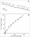
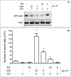
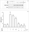
Binary toxins are among the most potent bacterial protein toxins performing a cooperative mode of translocation and exhibit fatal enzymatic activities in eukaryotic cells. Anthrax and C2 toxin are the most prominent examples for the AB(7/8) type of toxins. The B subunits bind both host cell receptors and the enzymatic A polypeptides to trigger their internalization and translocation into the host cell cytosol. C2 toxin is composed of an actin ADP-ribosyltransferase (C2I) and C2II binding subunits. Anthrax toxin is composed of adenylate cyclase (EF) and MAPKK protease (LF) enzymatic components associated to protective antigen (PA) binding subunit. The binding and translocation components anthrax protective antigen (PA(63)) and C2II of C2 toxin share a sequence homology of about 35%, suggesting that they might substitute for each other. Here we show by conducting in vitro measurements that PA(63) binds C2I and that C2II can bind both EF and LF. Anthrax edema factor (EF) and lethal factor (LF) have higher affinities to bind to channels formed by C2II than C2 toxin's C2I binds to anthrax protective antigen (PA(63)). Furthermore, we could demonstrate that PA in high concentration has the ability to transport the enzymatic moiety C2I into target cells, causing actin modification and cell rounding. In contrast, C2II does not show significant capacity to promote cell intoxication by EF and LF. Together, our data unveiled the remarkable flexibility of PA in promoting C2I heterologous polypeptide translocation into cells.
Conflict of interest statement
Figures


Similar articles
-
Role of N-terminal His6-Tags in binding and efficient translocation of polypeptides into cells using anthrax protective antigen (PA).PLoS One. 2012;7(10):e46964. doi: 10.1371/journal.pone.0046964. Epub 2012 Oct 8. PLoS One. 2012. PMID: 23056543 Free PMC article.
-
Structure, Function and Evolution of Clostridium botulinum C2 and C3 Toxins: Insight to Poultry and Veterinary Vaccines.Curr Protein Pept Sci. 2017;18(5):412-424. doi: 10.2174/1389203717666161201203311. Curr Protein Pept Sci. 2017. PMID: 27915984 Review.
-
Channel formation by the binding component of Clostridium botulinum C2 toxin: glutamate 307 of C2II affects channel properties in vitro and pH-dependent C2I translocation in vivo.Biochemistry. 2003 May 13;42(18):5368-77. doi: 10.1021/bi034199e. Biochemistry. 2003. PMID: 12731878
-
The host cell chaperone Hsp90 is essential for translocation of the binary Clostridium botulinum C2 toxin into the cytosol.J Biol Chem. 2003 Aug 22;278(34):32266-74. doi: 10.1074/jbc.M303980200. Epub 2003 Jun 12. J Biol Chem. 2003. PMID: 12805360
-
Clostridium botulinum C2 toxin--new insights into the cellular up-take of the actin-ADP-ribosylating toxin.Int J Med Microbiol. 2004 Apr;293(7-8):557-64. doi: 10.1078/1438-4221-00305. Int J Med Microbiol. 2004. PMID: 15149031 Review.
Cited by
-
Channel-forming bacterial toxins in biosensing and macromolecule delivery.Toxins (Basel). 2014 Aug 21;6(8):2483-540. doi: 10.3390/toxins6082483. Toxins (Basel). 2014. PMID: 25153255 Free PMC article. Review.
-
Role of N-terminal His6-Tags in binding and efficient translocation of polypeptides into cells using anthrax protective antigen (PA).PLoS One. 2012;7(10):e46964. doi: 10.1371/journal.pone.0046964. Epub 2012 Oct 8. PLoS One. 2012. PMID: 23056543 Free PMC article.
-
Obstructing toxin pathways by targeted pore blockage.Chem Rev. 2012 Dec 12;112(12):6388-430. doi: 10.1021/cr300141q. Epub 2012 Oct 11. Chem Rev. 2012. PMID: 23057504 Free PMC article. Review. No abstract available.
-
A human/murine chimeric fab antibody neutralizes anthrax lethal toxin in vitro.Clin Dev Immunol. 2013;2013:475809. doi: 10.1155/2013/475809. Epub 2013 Jun 5. Clin Dev Immunol. 2013. PMID: 23861692 Free PMC article.
-
Characterization and Pharmacological Inhibition of the Pore-Forming Clostridioides difficile CDTb Toxin.Toxins (Basel). 2021 May 28;13(6):390. doi: 10.3390/toxins13060390. Toxins (Basel). 2021. PMID: 34071730 Free PMC article.
References
-
- Collier RJ, Young JA. Anthrax toxin. Annual Review of Cell and Developmental Biology. 2003;19:45–70. - PubMed
-
- Young JA, Collier RJ. Anthrax toxin: receptor binding, internalization, pore formation, and translocation. Annu Rev Biochem. 2007;76:243–265. - PubMed
-
- Turk BE. Manipulation of host signalling pathways by anthrax toxins. Biochem J. 2007;402:405–417. - PubMed
-
- Rolando M, Stefani C, Flatau G, Auberger P, Mettouchi A, et al. Transcriptome dysregulation by anthrax lethal toxin plays a key role in induction of human endothelial cell cytotoxicity. Cell Microbiol. 2010;12:891–905. - PubMed
Publication types
MeSH terms
Substances
LinkOut - more resources
Full Text Sources
Miscellaneous

