Review
. 2011 Oct;51(1):45-60.
doi: 10.1007/s12026-011-8239-5.

Membrane attack by complement: the assembly and biology of terminal complement complexes

Affiliations
Review

Membrane attack by complement: the assembly and biology of terminal complement complexes

Cosmin A Tegla et al. Immunol Res. 2011 Oct.

Abstract

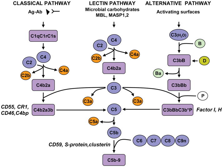
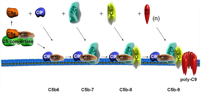
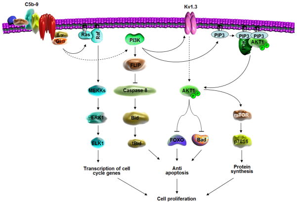
Complement system activation plays an important role in both innate and acquired immunity. Activation of the complement and the subsequent formation of C5b-9 channels (the membrane attack complex) on the cell membranes lead to cell death. However, when the number of channels assembled on the surface of nucleated cells is limited, sublytic C5b-9 can induce cell cycle progression by activating signal transduction pathways and transcription factors and inhibiting apoptosis. This induction by C5b-9 is dependent upon the activation of the phosphatidylinositol 3-kinase/Akt/FOXO1 and ERK1 pathways in a Gi protein-dependent manner. C5b-9 induces sequential activation of CDK4 and CDK2, enabling the G1/S-phase transition and cellular proliferation. In addition, it induces RGC-32, a novel gene that plays a role in cell cycle activation by interacting with Akt and the cyclin B1-CDC2 complex. C5b-9 also inhibits apoptosis by inducing the phosphorylation of Bad and blocking the activation of FLIP, caspase-8, and Bid cleavage. Thus, sublytic C5b-9 plays an important role in cell activation, proliferation, and differentiation, thereby contributing to the maintenance of cell and tissue homeostasis.


Figures

Fig. 1
Fig. 1. Complement activation pathways and assembly of the terminal pathway
The classical pathway is initiated by the binding of the C1 complex to antibody already bound to antigen, leading to the formation of the C4b2a enzyme complex (the C3 convertase). The lectin pathway is activated by the binding of either MBL or ficolin and MASP 1, 2, and 3, respectively, to an array of mannose groups on the surface of bacterial cells and the generation of C3 convertase of the classical pathway. The alternative pathway is initiated by hydrolyzed C3 and factor B and the subsequent formation of the alternative pathway C3 convertase, C3bBb. Generation of the C3 convertase allows the formation of the C5 convertase enzyme, which initiates the formation of the C5b-9 terminal complement complex. The complement system is regulated at several levels: CD55, CR1, CD46, C4bp, and factors I and H regulate the activity of the C3 convertase and C5 convertase, and other proteins such as CD59 block the final assembly of the pores by preventing the binding of C9. The S protein/vitronectin binds to C5b-7 and leads to the formation of a cytolytically inactive SC5b-9 complex
Fig. 2
Fig. 2. Assembly of terminal complement complex
The formation and assembly of C5 convertase occurs via either the classical, alternative, or mannose-binding lectin pathway. C5 convertase binds to C5 and cleaves it to generate C5a and C5b. After binding to C5b, C6 acquires the ability to interact with the lipid bilayer and form a C5b6 bimolecular complex. C7 and then C8 sequentially bind to C5b and further insert into the lipid bilayer, forming C5b-7 and C5b-8 complexes, respectively. One molecule of C9 binds to membrane-inserted C8, after which the polymerization of multiple C9 molecules occurs to form the C5b-9 complex
Fig. 3
Fig. 3. Structural organization of the terminal complement system proteins
Six different types of structural modules are common to proteins of the human terminal components: the membrane attack complex/perforin domain (MACPF), the LDL receptor module (A), the EGF receptor module (EF), the thrombospondin I module (TSPI), the short consensus repeat (TSPI), and the factor I module (FIM). Gray hexagonal symbols indicate the asparagine-linked glycosylation sites
Fig. 4
Fig. 4. Signaling pathways induced by C5b-9 that play a role in cellular proliferation and protection from apoptosis
C5b-9 interacts with the Gi protein, inducing activation of Ras. Ras then activates Raf-1, which in turn activates MEK1 and ERK1. C5b-9 also activates PI3K and induces the activation of PDK1 and Akt. PI3K also participates in the activation of ERK1 and p70S6 kinase, leading to the activation of the cell cycle. Kv1.3 is also involved in cell cycle activation and is induced by C5b-9 via the modulation of Akt activation. Activation of PI3K induces up-regulation of FLIP and inhibits caspase-8 processing. C5b-9-induced Akt activation also inhibits apoptosis by regulating FOXO1, Bad phosphorylation and inhibiting Bid cleavage

References

    1. Ehrlich P, Sachs H. Ueber die Vielheit der Complemente des Serums. Berliner Klinische Wochenschrift. 1902;14:297–338.
    1. Muller-Eberhard HJ. Molecular organization and function of the complement system. Annu Rev Biochem. 1988;57:321–347. - PubMed
    1. Walport MJ. Complement First of two parts. N Engl J Med. 2001;344:1058–1066. - PubMed
    1. Hugli TE. Biochemistry and biology of anaphylatoxins. Complement. 1986;3:111–127. - PubMed
    1. Frank MM. Complement disorders and hereditary angioedema. J Allergy Clin Immunol. 2010;125:S262–271. - PubMed

Publication types

MeSH terms

Substances