Review
. 2012 Jan;24(1):160-73.
doi: 10.1111/j.1365-2826.2011.02206.x.

Roles for oestrogen receptor β in adult brain function

Affiliations
Review

Roles for oestrogen receptor β in adult brain function

R J Handa et al. J Neuroendocrinol. 2012 Jan.

Abstract

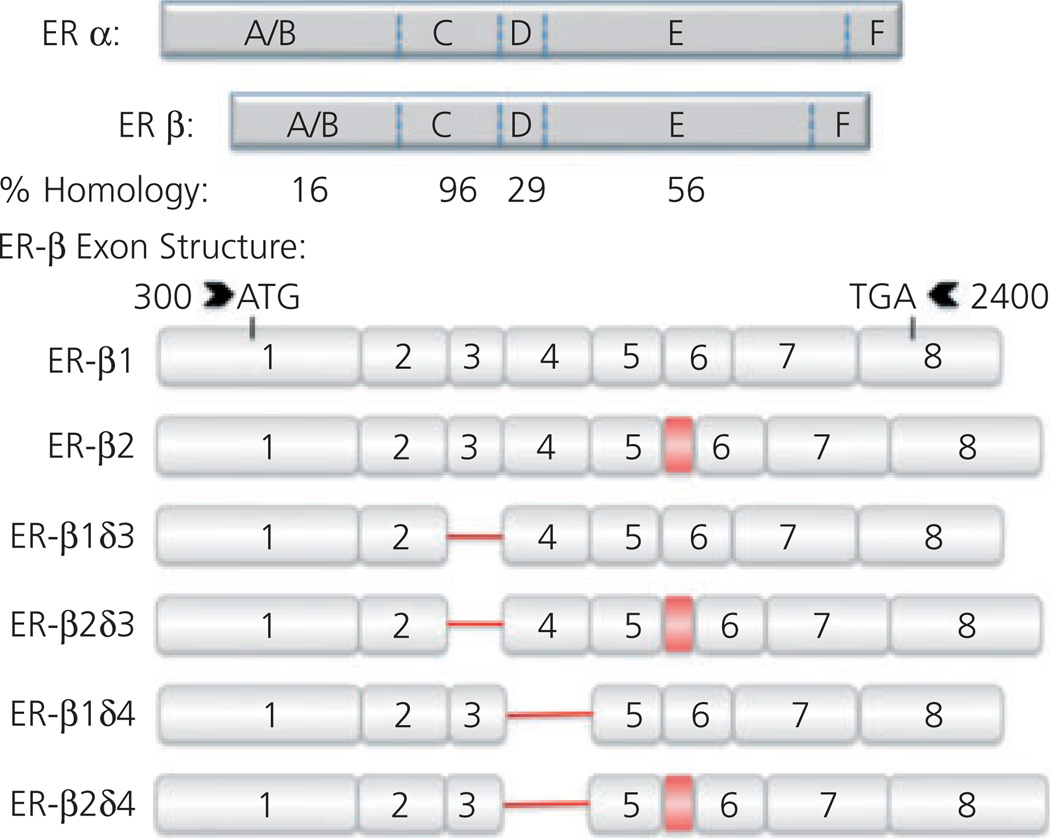
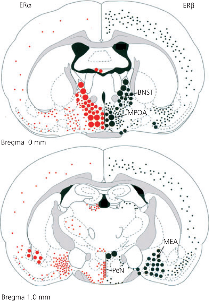
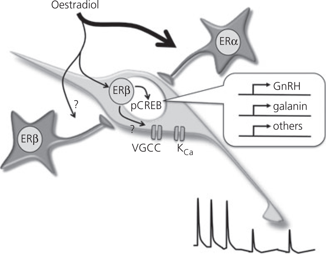
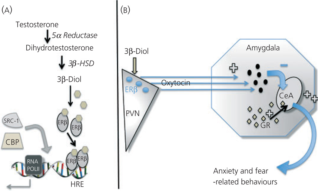
Oestradiol exerts a profound influence upon multiple brain circuits. For the most part, these effects are mediated by oestrogen receptor (ER)α. We review here the roles of ERβ, the other ER isoform, in mediating rodent oestradiol-regulated anxiety, aggressive and sexual behaviours, the control of gonadotrophin secretion, and adult neurogenesis. Evidence exists for: (i) ERβ located in the paraventricular nucleus underpinning the suppressive influence of oestradiol on the stress axis and anxiety-like behaviour; (ii) ERβ expressed in gonadotrophin-releasing hormone neurones contributing to oestrogen negative-feedback control of gonadotrophin secretion; (iii) ERβ controlling the offset of lordosis behaviour; (iv) ERβ suppressing aggressive behaviour in males; (v) ERβ modulating responses to social stimuli; and (vi) ERβ in controlling adult neurogenesis. This review highlights two major themes; first, ERβ and ERα are usually tightly inter-related in the oestradiol-dependent control of a particular brain function. For example, even though oestradiol feedback to control reproduction occurs principally through ERα-dependent mechanisms, modulatory roles for ERβ also exist. Second, the roles of ERα and ERβ within a particular neural network may be synergistic or antagonistic. Examples of the latter include the role of ERα to enhance, and ERβ to suppress, anxiety-like and aggressive behaviours. Splice variants such as ERβ2, acting as dominant negative receptors, are of further particular interest because their expression levels may reflect preceeding oestradiol exposure of relevance to oestradiol replacement therapy. Together, this review highlights the predominant modulatory, but nonetheless important, roles of ERβ in mediating the many effects of oestradiol upon adult brain function.


Figures

Fig. 1
Fig. 1
Oestrogen receptor (ER)α and ERβ homology and ERβ splice variants. Schematic representations of ERα and ERβ protein structure with relative percentage homology shown below. Letters refer to different domains of receptors; amino terminal domain (A/B), DNA-binding domain (C), a hinge region (D), ligand-binding domain (E) and caudal C-terminal (F). ERβ splice variant exon structure shown below. Exons 1–8 are numbered. Deletions are indicated by red line and insertions are indicated by a red box. The insertion between exons 5 and 6 (ERβ2) results in a modified ligand-binding domain (E). Splice variant data from J. M. Wang (unpublished data) and Price et al. (169).
Fig. 2
Fig. 2
Differential distribution of oestrogen receptor (ER) α and ERβ in the rodent brain. Two coronal planes through the brain (one at bregma and one at −1 mm to bregma for mouse) showing the anatomical distribution of ERα (left) and ERβ (right). Note the overlapping as well as differentially distributed expression of the two ERs. BNST, bed nucleus of the stria terminalis; MPOA, medial preoptic area; MEA, medial amygdala; PeN, periventricular nucleus. Grey shading shows white matter tracts. Adapted with permission (17).
Fig. 3
Fig. 3
Schematic diagram showing the various pathways underpinning oestrogen negative feedback in the rodent. The principal pathway involves indirect modulation of gonadotrophin-releasing hormone (GnRH) neurones through oestrogen receptor (ER)α. A second pathway that involves direct and indirect ERβ-dependent modulation is shown. Genes reported to be regulated by oestradiol through ERβ in GnRH neurones are shown in the inset. Within GnRH neurones, oestradiol is considered to modulate the phosphorylation status of cAMP response element binding (CREB) protein, providing another mechanism for transcriptional regulation. Two ion channels modulated by ERβ-dependent signalling within or outside the GnRH neurone are the voltage-gated calcium channels (VGCC) and calcium-activated potassium channels (KCa). Together, these pathways may help suppress pulsatile luteinising hormone secretion (bottom right).
Fig. 4
Fig. 4
Putative mechanisms for 5α-androstane-3β,17β-diol (3β-diol) inhibition of anxiety-like behaviours. (A) 3β-Diol is produced through oxidation of dihydrotestosterone by the enzyme, 3β-hydroxysteroid dehydrogenase (3β-HSD) and others. Within oxytocin (OT) neurones, it binds ERβ, which dimerises and activates OT gene transcription by binding the hormone response element located at −160 in the ot promoter. In doing so, it attracts co-regulatory proteins such as SRC1 and CBP to regulate transcription of the ot gene. (B) 3β-Diol binds and activates ERβ found in OT neurones of the paraventricular nucleus (PVN) to activate inhibitory neurones in the amygdala and correspondingly reduce activity of neurones in the central nucleus of the amygdala (CeA). The activation of CeA neurones is involved in increased anxiety- and fear-related behaviours. By contrast, glucocorticoid receptor (GR) containing neurones of the amygdala and CeA will increase the tone of CeA neurones, thereby potentiating fear- and anxiety-related behaviours. HRE, hormone response element

References

    1. Walter P, Green S, Greene G, Krust A, Bornert JM, Jeltsch JM, Staub A, Jensen E, Scrace G, Waterfield M, Chambon P. Cloning of the human estrogen receptor cDNA. Proc Natl Acad Sci USA. 1985;82:7889–7893. - PMC - PubMed
    1. Kuiper GG, Enmark E, Pelto-Huikko M, Nilsson S, Gustafsson JA. Cloning of a novel receptor expressed in rat prostate and ovary. Proc Natl Acad Sci USA. 1996;93:5925–5930. - PMC - PubMed
    1. Beato M, Herrlich P, Schutz G. Steroid hormone receptors: many actors in search of a plot. Cell. 1995;83:851–857. - PubMed
    1. Robinson-Rechavi M, Escriva Garcia H, Laudet V. The nuclear receptor superfamily. J Cell Sci. 2003;116:585–586. - PubMed
    1. Poola I, Abraham J, Baldwin K. Identification of ten exon deleted ERbeta mRNAs in human ovary, breast, uterus and bone tissues: alternate splicing pattern of estrogen receptor beta mRNA is distinct from that of estrogen receptor alpha. FEBS Lett. 2002;516:133–138. - PubMed

Publication types

Substances