Potato virus X movement in Nicotiana benthamiana: new details revealed by chimeric coat protein variants
- PMID: 21851552
- PMCID: PMC6638808
- DOI: 10.1111/j.1364-3703.2011.00739.x
Potato virus X movement in Nicotiana benthamiana: new details revealed by chimeric coat protein variants
Abstract
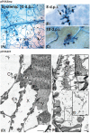
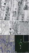
Potato virus X coat protein is necessary for both cell-to-cell and phloem transfer, but it has not been clarified definitively whether it is needed in both movement phases solely as a component of the assembled particles or also of differently structured ribonucleoprotein complexes. To clarify this issue, we studied the infection progression of a mutant carrying an N-terminal deletion of the coat protein, which was used to construct chimeric virus particles displaying peptides selectively affecting phloem transfer or cell-to-cell movement. Nicotiana benthamiana plants inoculated with expression vectors encoding the wild-type, mutant and chimeric viral genomes were examined by microscopy techniques. These experiments showed that coat protein-peptide fusions promoting cell-to-cell transfer only were not competent for virion assembly, whereas long-distance movement was possible only for coat proteins compatible with virus particle formation. Moreover, the ability of the assembled PVX to enter and persist into developing xylem elements was revealed here for the first time.
PUBLISHED 2011. THIS ARTICLE IS A US GOVERNMENT WORK AND IS IN THE PUBLIC DOMAIN IN THE USA.
Figures


Similar articles
-
Complementation of the movement-deficient mutations in potato virus X: potyvirus coat protein mediates cell-to-cell trafficking of C-terminal truncation but not deletion mutant of potexvirus coat protein.Virology. 2000 Apr 25;270(1):31-42. doi: 10.1006/viro.2000.0246. Virology. 2000. PMID: 10772977
-
Peptide display on Potato virus X: molecular features of the coat protein-fused peptide affecting cell-to-cell and phloem movement of chimeric virus particles.J Gen Virol. 2006 Oct;87(Pt 10):3103-3112. doi: 10.1099/vir.0.82097-0. J Gen Virol. 2006. PMID: 16963770
-
Identification of viral particles in the apoplast of Nicotiana benthamiana leaves infected by potato virus X.Mol Plant Pathol. 2021 Apr;22(4):456-464. doi: 10.1111/mpp.13039. Epub 2021 Feb 24. Mol Plant Pathol. 2021. PMID: 33629491 Free PMC article.
-
Potato virus X TGBp1 induces plasmodesmata gating and moves between cells in several host species whereas CP moves only in N. benthamiana leaves.Virology. 2004 Oct 25;328(2):185-97. doi: 10.1016/j.virol.2004.06.039. Virology. 2004. PMID: 15464839
-
Viral and nonviral elements in potexvirus replication and movement and in antiviral responses.Adv Virus Res. 2013;87:75-112. doi: 10.1016/B978-0-12-407698-3.00003-X. Adv Virus Res. 2013. PMID: 23809921 Review.
Cited by
-
Sequence-Specific Protein Aggregation Generates Defined Protein Knockdowns in Plants.Plant Physiol. 2016 Jun;171(2):773-87. doi: 10.1104/pp.16.00335. Epub 2016 May 4. Plant Physiol. 2016. PMID: 27208282 Free PMC article.
-
A GoldenBraid-Compatible Virus-Based Vector System for Transient Expression of Heterologous Proteins in Plants.Viruses. 2022 May 20;14(5):1099. doi: 10.3390/v14051099. Viruses. 2022. PMID: 35632840 Free PMC article.
-
Atomic structure of potato virus X, the prototype of the Alphaflexiviridae family.Nat Chem Biol. 2020 May;16(5):564-569. doi: 10.1038/s41589-020-0502-4. Epub 2020 Mar 16. Nat Chem Biol. 2020. PMID: 32203412
-
Blaze a New Trail: Plant Virus Xylem Exploitation.Int J Mol Sci. 2022 Jul 29;23(15):8375. doi: 10.3390/ijms23158375. Int J Mol Sci. 2022. PMID: 35955508 Free PMC article. Review.
-
The Two-Faced Potato Virus X: From Plant Pathogen to Smart Nanoparticle.Front Plant Sci. 2015 Nov 17;6:1009. doi: 10.3389/fpls.2015.01009. eCollection 2015. Front Plant Sci. 2015. PMID: 26635836 Free PMC article. Review.
References
-
- Atabekov, J. , Dobrov, E. , Karpova, O. and Radionova, N. (2007) Potato virus X: structure, disassembly and reconstitution. Mol. Plant. Pathol. 8, 667–675. - PubMed
-
- Atabekov, J.G. , Rodionova, N.P. , Karpova, O.V. , Kozlovsky, S.V. , Novikov, V.K. and Arkhipenko, M.V. (2001) Translational activation of encapsidated Potato virus X RNA by coat protein phosphorylation. Virology, 286, 466–474. - PubMed
-
- Bamunusinghe, D. , Hemenway, C.L. , Nelson, R.S. , Sanderfoot, A.A. , Ye, C.M. , Silva, M.A.T. , Payton, M. and Verchot‐Lubicz, J. (2009) Analysis of potato virus X replicase and TGBp3 subcellular locations. Virology, 393, 272–285. - PubMed
-
- Baratova, L.A. , Fedorova, N.V. , Dobrov, E.N. , Lukashina, E.V. , Kharlanov, A.N. , Nasonov, V.V. , Serebryakova, M.V. , Kozlovsky, S.V. , Zayakina, O.V. and Rodionova, N.P. (2004) N‐terminal segment of Potato virus X coat protein subunits is glycosylated and mediates formation of a bound water shell on the virion surface. Eur. J. Biochem. 271, 3136–3145. - PubMed
-
- Baulcombe, D.C. , Chapman, S. and Santa Cruz, S. (1995) Jellyfish green fluorescent protein as a reporter for virus infections. Plant J. 7, 1045–1053. - PubMed
Publication types
MeSH terms
Substances
LinkOut - more resources
Full Text Sources

