. 2011 Aug 18:8:102.
doi: 10.1186/1742-2094-8-102.

Anandamide inhibits Theiler's virus induced VCAM-1 in brain endothelial cells and reduces leukocyte transmigration in a model of blood brain barrier by activation of CB(1) receptors

Affiliations

Anandamide inhibits Theiler's virus induced VCAM-1 in brain endothelial cells and reduces leukocyte transmigration in a model of blood brain barrier by activation of CB(1) receptors

Leyre Mestre et al. J Neuroinflammation. .

Abstract

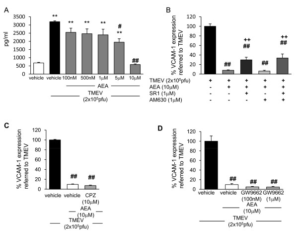
Background: VCAM-1 represents one of the most important adhesion molecule involved in the transmigration of blood leukocytes across the blood-brain barrier (BBB) that is an essential step in the pathogenesis of MS. Several evidences have suggested the potential therapeutic value of cannabinoids (CBs) in the treatment of MS and their experimental models. However, the effects of endocannabinoids on VCAM-1 regulation are poorly understood. In the present study we investigated the effects of anandamide (AEA) in the regulation of VCAM-1 expression induced by Theiler's virus (TMEV) infection of brain endothelial cells using in vitro and in vivo approaches.

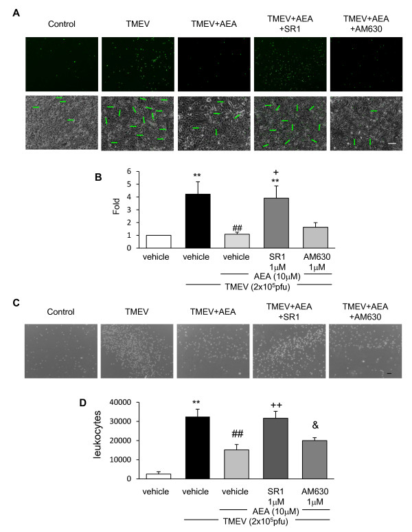
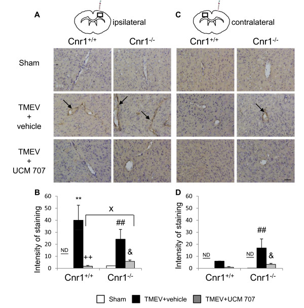
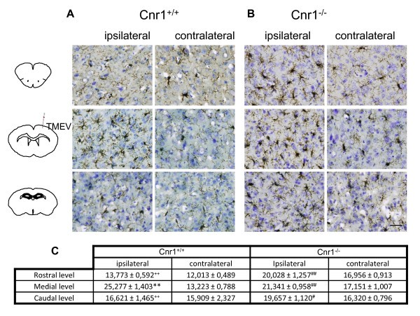
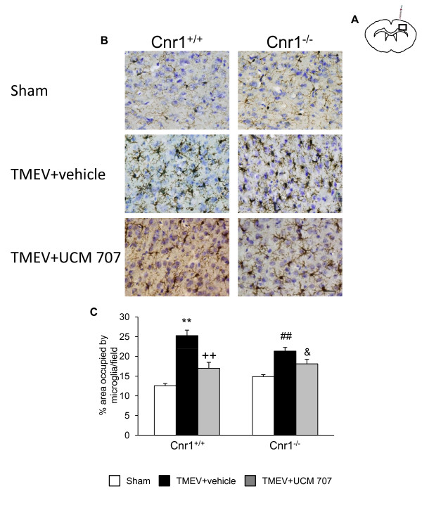
Methods: i) in vitro: VCAM-1 was measured by ELISA in supernatants of brain endothelial cells infected with TMEV and subjected to AEA and/or cannabinoid receptors antagonist treatment. To evaluate the functional effect of VCAM-1 modulation we developed a blood brain barrier model based on a system of astrocytes and brain endothelial cells co-culture. ii) in vivo: CB(1) receptor deficient mice (Cnr1(-/-)) infected with TMEV were treated with the AEA uptake inhibitor UCM-707 for three days. VCAM-1 expression and microglial reactivity were evaluated by immunohistochemistry.

Results: Anandamide-induced inhibition of VCAM-1 expression in brain endothelial cell cultures was mediated by activation of CB(1) receptors. The study of leukocyte transmigration confirmed the functional relevance of VCAM-1 inhibition by AEA. In vivo approaches also showed that the inhibition of AEA uptake reduced the expression of brain VCAM-1 in response to TMEV infection. Although a decreased expression of VCAM-1 by UCM-707 was observed in both, wild type and CB(1) receptor deficient mice (Cnr1(-/-)), the magnitude of VCAM-1 inhibition was significantly higher in the wild type mice. Interestingly, Cnr1(-/-) mice showed enhanced microglial reactivity and VCAM-1 expression following TMEV infection, indicating that the lack of CB(1) receptor exacerbated neuroinflammation.

Conclusions: Our results suggest that CB(1) receptor dependent VCAM-1 inhibition is a novel mechanism for AEA-reduced leukocyte transmigration and contribute to a better understanding of the mechanisms underlying the beneficial role of endocannabinoid system in the Theiler's virus model of MS.


Figures

Figure 1
Figure 1
Anandamide inhibits VCAM-1 production induced by TMEV through a mechanism that involves CB1 receptor. (A) sVCAM-1 levels were measured by ELISA in supernatants of cell cultures 20 h after AEA treatment (100 nM, 500 nM, 1 μM, 5 μM, 10 μM). Confluent TMEV-infected brain endothelial cell monolayers were pretreated for 1 hour before AEA treatment with (B) the cannabinoid receptor antagonist SR1 (1 μM) or AM630 (1 μM); (C) the vanilloid receptor antagonist capsazepine (10 μM); (D) the PPARγ receptor antagonist GW9662 (100 nM and 1 μM). Results show the means ± SEM from three independent experiments done in triplicate. (**p < 0,01 vs. vehicle; ##p < 0.01 vs. TMEV+vehicle; ++p < 0.01 vs. TMEV+AEA, ANOVA followed by Tuckey's test).
Figure 2
Figure 2
AEA limits leukocyte adhesion to TMEV stimulated brain endothelial cells and leukocyte transmigration through in vitro BBB by CB1 involvement. Brain endothelial cell monolayer were stimulated with a combination of TMEV (2 × 10⁵ pfu), AEA (10 μM), SR1 (1 μM) or AM630 (1 μM) for 6 hours. After that, 2.5 × 10⁵ leukocytes stained with AM-calcein (5 μM) were added to the endothelial culture for 20 hours. (A) Representative immunofluorescence microphotographs of the leukocytes stained with AM-calcein adhered to the brain endothelial cell monolayer in each case and phase contrast microphotographs of brain endothelial monolayer merged with immunofluorescence microphotographs of AM-calcein stained leukocytes bring out with arrows. Scale bar 100 μm. (B) Quantification of leukocytes adhered to brain endothelial monolayer in each case normalized to control group (n = 6). (**p < 0.01 vs. vehicle; ##p < 0.01 vs. TMEV; +p < 0.05 vs. TMEV+AEA, ANOVA followed by Tuckey's tests). (C) TMEV (2 × 10⁵ pfu), plus AEA (10 μM), or plus SR1 (1 μM) or AM630 (1 μM) were added to the upper side of the insert (endothelial culture) and IL1-β (10 ng/ml) was added to the bottom side (astrocyte culture) for 6 hours. 2.5 × 10⁵ leukocytes were added to the upper side of the insert for 20 hours and representative phase contrast microphotographs of leukocytes crossed to bottom side of the insert were taken. (D) Quantification of leukocytes in the bottom side of the insert after 20 hours of experiment. (**p < 0.01 vs. vehicle; ##p < 0.01 vs. TMEV+vehicle; ++p < 0.01 vs. TMEV+AEA; &p < 0.05 vs. TMEV+AEA+SR1, ANOVA followed by Tuckey's test; n = 6).
Figure 3
Figure 3
The treatment with UCM-707 inhibits VCAM-1 expression in TMEV-infected mice. Study with Cnr1+/+ and Cnr1-/- mice. Both, TMEV-infected and Sham mice were treated with UCM-707 (3 mg/kg) or the corresponding vehicle (n = 3 for each group) immediately after virus infection for three consecutive days. Analysis were performed using representative microphotographs of coronal brain sections (30 μm) of ipsilateral (A) or contralateral (C) brain tissue close to the virus side of injection, immunostained for VCAM-1. Arrows indicate VCAM-1 immunostaining. Scale bar is 50 μm. (B, D) Quantification of intensity of VCAM-1 staining as described in Material and methods in the ipsilateral or contralateral hemispheres, respectively. ND, non detected; **p < 0.01 vs. Sham (Cnr1+/+); ##p < 0.01 vs. Sham (Cnr1-/-); ++p < 0.01 vs. TMEV+vehicle (Cnr1+/+); &p < 0.05 vs. TMEV+vehicle (Cnr1-/-); Xp < 0.05 vs. TMEV+UCM-707 (Cnr1+/+).
Figure 4
Figure 4
CB1 deletion exacerbates microglial response against TMEV infection. Coronal brain sections (30 μm) were obtained from Cnr1+/+ TMEV-infected mice (A) or Cnr1-/- TMEV-infected mice (B), stained for CD11b with Iba-1 antibody and counterstained with toluidine blue (n = 3 for each group). To perform the analysis of microglia phenotype morphology brain tissue was studied in both hemispheres and at rostral, medial and caudal levels. (C) Quantification of percentage of area occupied by microglia per field is represented. Scale bar is 50 μm. **p < 0.01 vs. contralateral (Cnr1+/+); #p < 0.05 vs. contralateral (Cnr1-/-); ##p < 0.01 vs. contralateral (Cnr1-/-); ++p < 0.01 vs. medial level (Cnr1+/+).
Figure 5
Figure 5
The treatment with UCM-707 decreases microglia reactivity in TMEV-infected mice. Study with Cnr1+/+ and Cnr1-/- mice. Both TMEV-infected and Sham mice from both strains (Cnr1+/+ and Cnr1-/-) were treated with UCM-707 (3 mg/kg) or the corresponding vehicle (n = 3 for each group) immediately after the virus infection for three consecutive days. (A) Coronal brain section level for the analysis of CD11b+ expression. (B) Representative micrographs of ipsilateral cerebral cortex in sham, TMEV-infected plus vehicle or TMEV infected plus UCM-707 from Cnr1+/+ or Cnr1-/- mice. (C) Quantification of percentage of area occupied by microglia per field is represented. Scale bar is 50 μm. **p < 0.01 vs Sham (Cnr1+/+); ++p < 0.01 vs. TMEV+vehicle (Cnr1+/+); ##p < 0.01 vs Sham (Cnr1-/-); &p = 0.07 vs. TMEV+vehicle (Cnr1-/-).

Similar articles

Cited by

References

    1. Engelhardt B, Ransohoff RM. The ins and outs of T-lymphocyte trafficking to the CNS: anatomical sites and molecular mechanisms. Trends Immunol. 2005;26:485–495. doi: 10.1016/j.it.2005.07.004. - DOI - PubMed
    1. Greenwood J, Heasman SJ, Alvarez JI, Prat A, Lyck R, Engelhardt B. Leucocyte-endothelial cell crosstalk at the blood-brain barrier: A prerequisite for successful immune cell entry to the brain. Neuropathol Appl Neurobiol. 2011;37:24–39. doi: 10.1111/j.1365-2990.2010.01140.x. - DOI - PubMed
    1. Ukkonen M, Wu K, Reipert B, Dastidar P, Elovaara I. Cell surface adhesion molecules and cytokine profiles in primary progressive multiple sclerosis. Mult Scler. 2007;13:701–707. doi: 10.1177/1352458506075378. - DOI - PubMed
    1. Brosnan CF, Cannella B, Battistini L, Raine CS. Cytokine localization in multiple sclerosis lesions: correlation with adhesion molecule expression and reactive nitrogen species. Neurology. 1995;45:S16–S21. - PubMed
    1. Yednock TA, Cannon C, Fritz LC, Sanchez-Madrid F, Steinman L, Karin N. Prevention of experimental autoimmune encephalomyelitis by antibodies against alpha 4 beta 1 integrin. Nature. 1992;356:63–66. doi: 10.1038/356063a0. - DOI - PubMed

Publication types

MeSH terms