

Transcriptome profiling of sheep granulosa cells and oocytes during early follicular development obtained by laser capture microdissection
- PMID: 21851638
- PMCID: PMC3166951
- DOI: 10.1186/1471-2164-12-417
Transcriptome profiling of sheep granulosa cells and oocytes during early follicular development obtained by laser capture microdissection
Abstract
Background: Successful achievement of early folliculogenesis is crucial for female reproductive function. The process is finely regulated by cell-cell interactions and by the coordinated expression of genes in both the oocyte and in granulosa cells. Despite many studies, little is known about the cell-specific gene expression driving early folliculogenesis. The very small size of these follicles and the mixture of types of follicles within the developing ovary make the experimental study of isolated follicular components very difficult.The recently developed laser capture microdissection (LCM) technique coupled with microarray experiments is a promising way to address the molecular profile of pure cell populations. However, one main challenge was to preserve the RNA quality during the isolation of single cells or groups of cells and also to obtain sufficient amounts of RNA.Using a new LCM method, we describe here the separate expression profiles of oocytes and follicular cells during the first stages of sheep folliculogenesis.
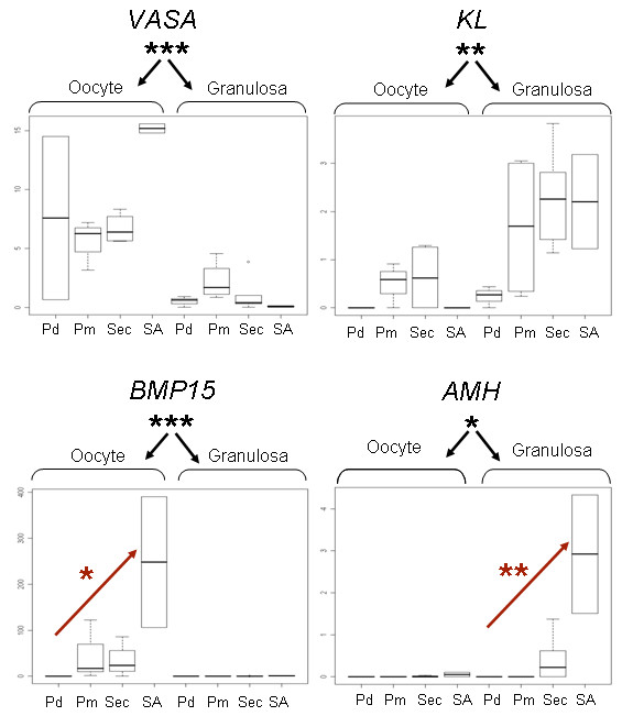
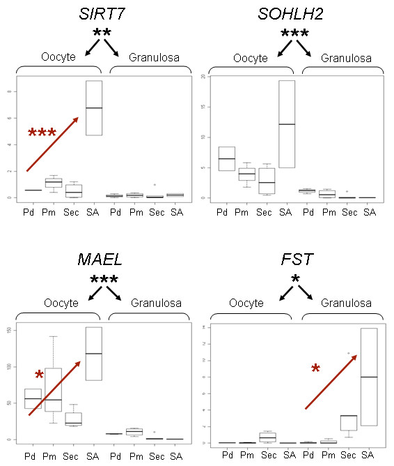
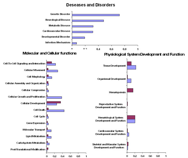
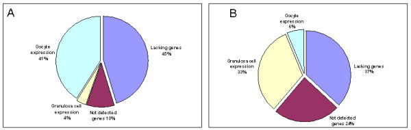
Results: We developed a new tissue fixation protocol ensuring efficient single cell capture and RNA integrity during the microdissection procedure. Enrichment in specific cell types was controlled by qRT-PCR analysis of known genes: six oocyte-specific genes (SOHLH2, MAEL, MATER, VASA, GDF9, BMP15) and three granulosa cell-specific genes (KL, GATA4, AMH).A global gene expression profile for each follicular compartment during early developmental stages was identified here for the first time, using a bovine Affymetrix chip. Most notably, the granulosa cell dataset is unique to date. The comparison of oocyte vs. follicular cell transcriptomes revealed 1050 transcripts specific to the granulosa cell and 759 specific to the oocyte.Functional analyses allowed the characterization of the three main cellular events involved in early folliculogenesis and confirmed the relevance and potential of LCM-derived RNA.
Conclusions: The ovary is a complex mixture of different cell types. Distinct cell populations need therefore to be analyzed for a better understanding of their potential interactions. LCM and microarray analysis allowed us to identify novel gene expression patterns in follicular cells at different stages and in oocyte populations.
Figures
References
-
- McNatty KP, Smith P, Hudson NL, Heath DA, Tisdall DJ, O WS, Braw-Tal R. Development of the sheep ovary during fetal and early neonatal life and the effect of fecundity genes. J Reprod Fertil Suppl. 1995;49:123–135. - PubMed
-
- Soyal SM, Amleh A, Dean J. FIGalpha, a germ cell-specific transcription factor required for ovarian follicle formation. Development. 2000;127:4645–4654. - PubMed
Publication types
MeSH terms
Substances
LinkOut - more resources
Full Text Sources
Other Literature Sources
Molecular Biology Databases
