. 2011 Nov;63(14-15):1282-99.
doi: 10.1016/j.addr.2011.07.001. Epub 2011 Aug 6.

Hybrid magnetic nanostructures (MNS) for magnetic resonance imaging applications

Affiliations

Hybrid magnetic nanostructures (MNS) for magnetic resonance imaging applications

Mrinmoy De et al. Adv Drug Deliv Rev. 2011 Nov.

Abstract

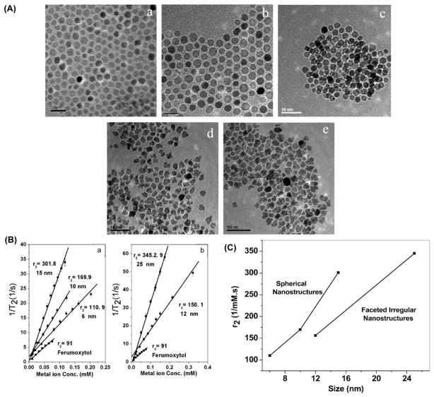
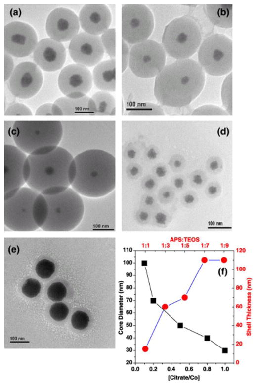
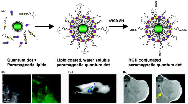
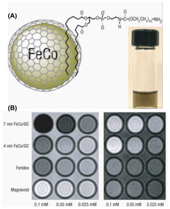
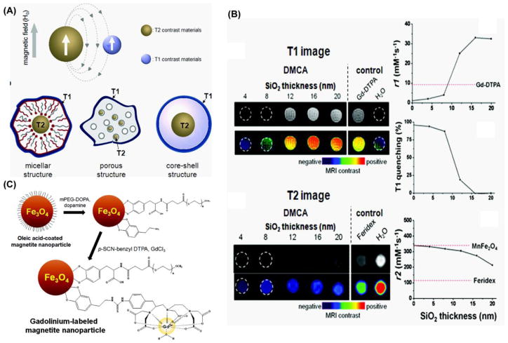
The development of MRI contrast agents has experienced its version of the gilded age over the past decade, thanks largely to the rapid advances in nanotechnology. In addition to progress in single mode contrast agents, which ushered in unprecedented R(1) or R(2) sensitivities, there has also been a boon in the development of agents covering more than one mode of detection. These include T(1)-PET, T(2)-PET T(1)-optical, T(2)-optical, T(1)-T(2) agents and many others. In this review, we describe four areas which we feel have experienced particular growth due to nanotechnology, specifically T(2) magnetic nanostructure development, T(1)/T(2)-optical dual mode agents, and most recently the T(1)-T(2) hybrid imaging systems. In each of these systems, we describe applications including in vitro, in vivo usage and assay development. In all, while the benefits and drawbacks of most MRI contrast agents depend on the application at hand, the recent development in multimodal nanohybrids may curtail the shortcomings of single mode agents in diagnostic and clinical settings by synergistically incorporating functionality. It is hoped that as nanotechnology advances over the next decade, it will produce agents with increased diagnostics and assay relevant capabilities in streamlined packages that can meaningfully improve patient care and prognostics. In this review article, we focus on T(2) materials, its surface functionalization and coupling with optical and/or T(1) agents.


Figures

Figure 1
Figure 1
Nanohybrid based various MR imaging agents.
Figure 2
Figure 2
MR properties of MNS is dependent on shape and size. (A) Synthesis results of various CoFe2O4 based on size, and shape regularity. It can be seen that pictures in A.a,b,c are spherical in nature, while A.d,e are faceted irregular (FI). (B). Relaxivity properties of MNS. (C). FI MNS demonstrate lower R2 values for a given size (as measured by longest diameter across). [30]
Figure 3
Figure 3
Co MNS can be controllably encapsulated in silica shell by manipulation of TEOS precursor ratio. Additionally, core diameter can be manipulated through capping ligand addition [38].
Figure 4
Figure 4
(A) Stabilization of iron oxide MNS using various catechol derivative as anchor group. (B) DLS measurements of individually stabilized iron oxide nanoparticles based on their aqueous stability (a), effect on filtration (b) and the stability as a function of temperature (c). (C) The dispersants ligands absorbed on 2D and nanoparticle surface as measured by XPS.
Figure 5
Figure 5
(A) Synthetic procedure of low and high pay load hybrid silica nanoparticle with [Ru(bpy)3]Cl2 and Gd3+. (B) TEM of above synthesized nanoparticles, showing the larger surface area of six times higher loaded gadolinium ion. The scale bar represents 200 nm (left) and 100 nm respectively. (C) Microscopic images of hybrid silica nanoparticles labeled monocyte cells: a) optical; b) laser scanning confocal fluorescence. c), d) MR images of unlabeled (left) and 1-labeled monocyte cells: c) T1-weighted and d) T2-weighted [92].
Figure 6
Figure 6
(A) Gd3+-DOTA functionalized CdSeTe/CdS QDs with glutathione (GSH) coating. (B) A NIR-fluorescence image [42] and T1 weighted MR image (bottom) of a mouse. A phantom containing 10 μM of Gd3+-DOTA-QDs was visualized by both fluorescence imaging and MRI. (C) QD with the biotinylated Gd-wedge, containing eight Gd-DTPA complexes with AnaxA5 (AnxA5-QD-Gd-wedge). This nanohybrid can be visualized by both fluorescence and MR imaging. (D) Schematic representation of a cNGR-labeled paramagnetic quantum dot. Commercially available this QD carries ~10 streptavidin moieties to which 6 cNGR groups and hence 24 gadolinium constructs were bound. Based on that estimation the maximum number of Gd3+ was 192 [–104].
Figure 7
Figure 7
(A) Schematic depiction of the preparation of liposome with a paramagnetic micellular coating and targeting peptide. (B) The brightfield (left) and fluorescence image of tumor-bearing mice after intravenous injection of paramagnetic QDs. (C) Fluorescence image reveled the target specific accumulation of contrast agents. (D) T1-weighted MR images before and 45 min after the injection of paramagnetic QD–micelles[106].
Figure 8
Figure 8
(A) Structure of Gd3+ chelating lipid Gd-DTPA-DSA and fluorescein labeled apolipoprotein E derived lipopeptide, P2fA2. (B–D) Confocal and MRI of untreated (B), incubated with 33% (C) and (D) 50% P2fA2 nanohybrid macrophage cell pellets. Scale bar: 20 μm [113].
Figure 9
Figure 9
Schematic presentation of various silica based optical-MNS nanohybrids. (A) Fluorophore doped MNS-Silica core-shell nanohybrids, (B) Fluorophore conjugated MNS-Silica core-shell nanohybrids and (C) MNS conjugated fluorophore doped silica nanohybrids.
Figure 10
Figure 10
(A) Schematic depiction of the synthetic procedure for Fe3O4-MSN (mesoporous silica nanoparticle), stabilization by PEG and loading of DOX (doxorubicin). (B,C) In vitro multimodal imaging of drug loaded MNS-silica nanohybrids, (B) fluorescence image (C) MRI image at different concentrations [131].
Figure 11
Figure 11
(A) Schematic diagram of phospholipid stabilized FeCo/graphite shell nanocrystal and a PBS suspension after heating to 80 °C for 1 h (inset). (B) T2 (left) and T1 weighted MR images of various contrast agents at three metal concentrations [50].
Figure 12
Figure 12
(A) Magnetic coupling between T1 and T2 contrast materials and the possible structures of various T1/T2 bimodal MRI contrast agent. (B) T1 and T2 MR imaging of MnFe2O4-Gd MNS with a variable thickness of SiO2 shell. With increasing SiO2 layer R1 increases and R2 decreases. (C) Synthesis of gadolinium-labeled MNS as a dual contrast agent for T1 and T2 weighted magnetic resonance imaging [148].

Similar articles

Cited by

References

    1. Brenner DJ, Hall EJ. Current concepts - Computed tomography - An increasing source of radiation exposure. New England Journal of Medicine. 2007;357:2277–2284. - PubMed
    1. Basu S, Chryssikos T, Moghadam-Kia S, Zhuang HM, Torigian DA, Alavi A. Positron Emission Tomography as a Diagnostic Tool in Infection: Present Role and Future Possibilities. Seminars in Nuclear Medicine. 2009;39:36–51. - PubMed
    1. Meerwaldt R, Slart RHJA, van Dam GM, Luijckx GJ, Tio RA, Zeebregts CJ. PET/SPECT imaging: From carotid vulnerability to brain viability. European Journal of Radiology. 2010;74:104–109. - PubMed
    1. Hong R, Fischer NO, Emrick T, Rotello VM. Surface PEGylation and Ligand Exchange Chemistry of FePt Nanoparticles for Biological Applications. Chemistry of Materials. 2005;17:4617–4621.
    1. Heeger DJ, Ress D. What does fMRI tell us about neuronal activity? Nature Reviews Neuroscience. 2002;3:142–151. - PubMed

Publication types