

Epicardial adipose tissue: emerging physiological, pathophysiological and clinical features
- PMID: 21852149
- PMCID: PMC4978122
- DOI: 10.1016/j.tem.2011.07.003
Epicardial adipose tissue: emerging physiological, pathophysiological and clinical features
Abstract
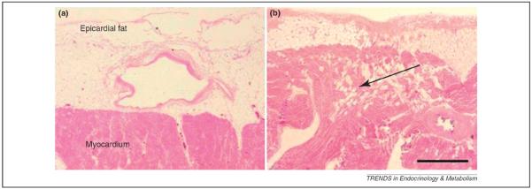
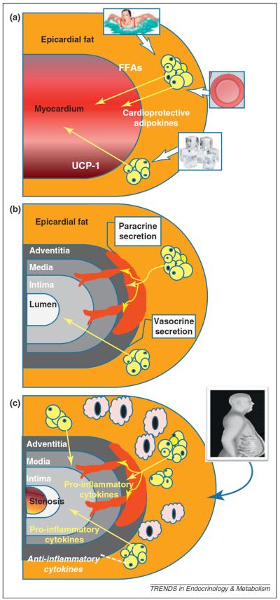
Epicardial adipose tissue is an unusual visceral fat depot with anatomical and functional contiguity to the myocardium and coronary arteries. Under physiological conditions, epicardial adipose tissue displays biochemical, mechanical and thermogenic cardioprotective properties. Under pathological circumstances, epicardial fat can locally affect the heart and coronary arteries through vasocrine or paracrine secretion of proinflammatory cytokines. What influences this equilibrium remains unclear. Improved local vascularization, weight loss, and targeted pharmaceutical interventions could help to return epicardial fat to its physiological role. This review focuses on the emerging physiological and pathophysiological aspects of the epicardial fat and its numerous and innovative clinical applications. Particular emphasis is placed on the paracrine/endocrine properties of epicardial fat and its role in the development and progression of atherosclerosis.
Copyright © 2011 Elsevier Ltd. All rights reserved.
Figures
Similar articles
-
Epicardial fat: from the biomolecular aspects to the clinical practice.Int J Biochem Cell Biol. 2011 Dec;43(12):1651-4. doi: 10.1016/j.biocel.2011.09.006. Epub 2011 Sep 28. Int J Biochem Cell Biol. 2011. PMID: 21967993 Review.
-
Epicardial adipose tissue and cardiac lipotoxicity: A review.Life Sci. 2023 Sep 1;328:121913. doi: 10.1016/j.lfs.2023.121913. Epub 2023 Jul 4. Life Sci. 2023. Retraction in: Life Sci. 2025 Jul 1;372:123630. doi: 10.1016/j.lfs.2025.123630. PMID: 37414140 Retracted. Review.
-
Epicardial Fat in the Maintenance of Cardiovascular Health.Methodist Debakey Cardiovasc J. 2017 Jan-Mar;13(1):20-24. doi: 10.14797/mdcj-13-1-20. Methodist Debakey Cardiovasc J. 2017. PMID: 28413578 Free PMC article. Review.
-
Local and systemic effects of the multifaceted epicardial adipose tissue depot.Nat Rev Endocrinol. 2015 Jun;11(6):363-71. doi: 10.1038/nrendo.2015.58. Epub 2015 Apr 7. Nat Rev Endocrinol. 2015. PMID: 25850659 Review.
-
Epicardial adipose tissue in endocrine and metabolic diseases.Endocrine. 2014 May;46(1):8-15. doi: 10.1007/s12020-013-0099-4. Epub 2013 Nov 23. Endocrine. 2014. PMID: 24272604 Review.
Cited by
-
Quantification of epicardial fat: Which method can predict significant coronary artery disease?World J Cardiol. 2015 May 26;7(5):287-92. doi: 10.4330/wjc.v7.i5.287. World J Cardiol. 2015. PMID: 26015859 Free PMC article.
-
Correlation analysis of epicardial adipose tissue and ventricular myocardial strain in Chinese amateur marathoners using cardiac magnetic resonance.PLoS One. 2022 Sep 13;17(9):e0274533. doi: 10.1371/journal.pone.0274533. eCollection 2022. PLoS One. 2022. PMID: 36099274 Free PMC article.
-
The Norwegian Stroke in the Young Study (NOR-SYS): rationale and design.BMC Neurol. 2013 Jul 17;13:89. doi: 10.1186/1471-2377-13-89. BMC Neurol. 2013. PMID: 23865483 Free PMC article.
-
An increase in epicardial adipose tissue is strongly associated with carotid-intima media thickness and atherosclerotic plaque, but LDL only with the plaque.Anatol J Cardiol. 2017 Jan;17(1):56-63. doi: 10.14744/AnatolJCardiol.2016.6885. Epub 2016 Aug 23. Anatol J Cardiol. 2017. PMID: 27564776 Free PMC article.
-
How do we measure epicardial adipose tissue thickness by transthoracic echocardiography?Anatol J Cardiol. 2015 May;15(5):416-9. doi: 10.5152/akd.2015.5991. Anatol J Cardiol. 2015. PMID: 25993714 Free PMC article. Review.
References
-
- Iacobellis G, et al. Epicardial adipose tissue: anatomic, biomolecular and clinical relationships with the heart. Nat. Clin. Pract. Cardiovasc. Med. 2005;2:536–543. - PubMed
-
- Iacobellis G. Epicardial and pericardial fat: close, but very different. Obesity. 2009;17:625. - PubMed
-
- Sacks HS, Fain JN. Human epicardial adipose tissue: a review. Am. Heart J. 2007;153:907–917. - PubMed
-
- Rabkin RW. Epicardial fat: properties, function and relationship to obesity. Obes. Rev. 2007;8:253–261. - PubMed
-
- Corradi D, et al. The ventricular epicardial fat is related to the myocardial mass in normal, ischemic and hypertrophic hearts. Cardiovasc. Pathol. 2004;13:313–316. - PubMed
Publication types
MeSH terms
Grants and funding
LinkOut - more resources
Full Text Sources
