. 2011 Oct 14;286(41):35522-35534.
doi: 10.1074/jbc.M111.274811. Epub 2011 Aug 18.

Identification and functional and spectral characterization of a globin-coupled histidine kinase from Anaeromyxobacter sp. Fw109-5

Affiliations

Identification and functional and spectral characterization of a globin-coupled histidine kinase from Anaeromyxobacter sp. Fw109-5

Kenichi Kitanishi et al. J Biol Chem. .

Abstract

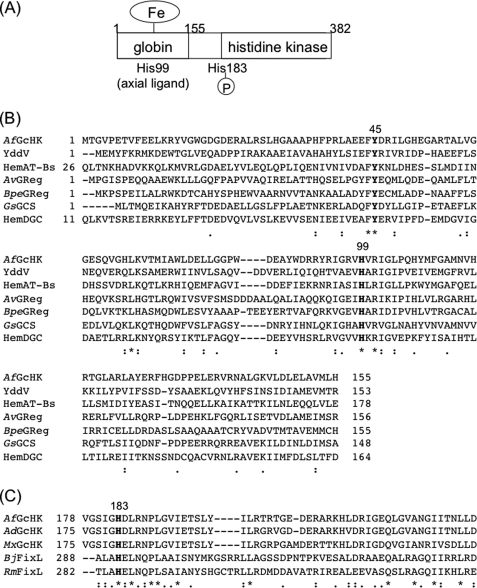
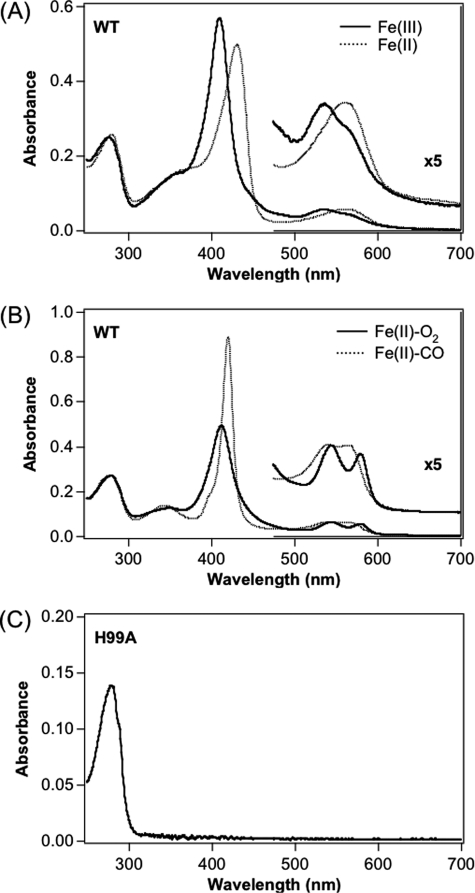
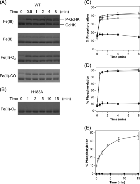
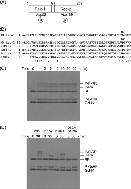
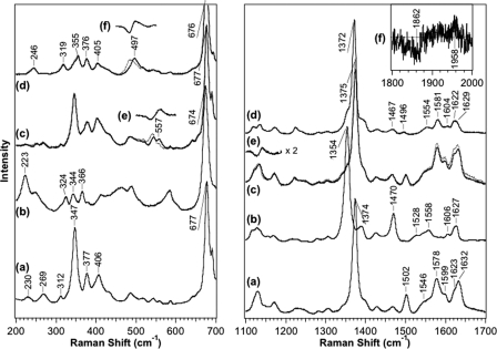
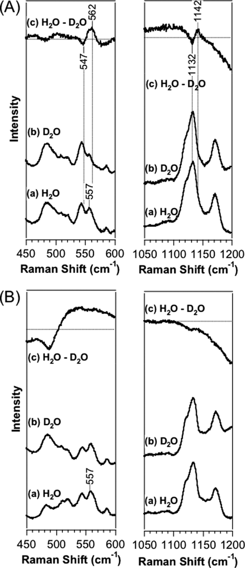
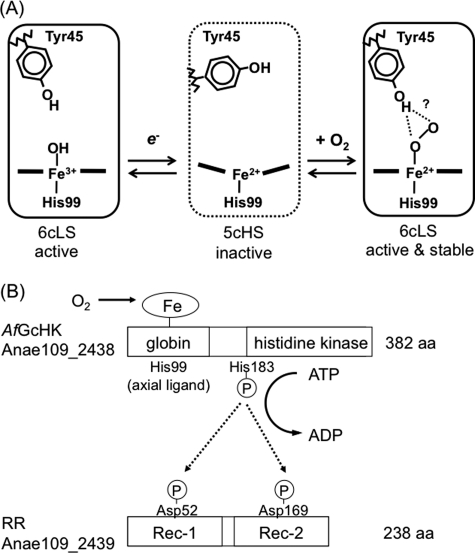
Two-component signal transduction systems regulate numerous important physiological functions in bacteria. In this study we have identified, cloned, overexpressed, and characterized a dimeric full-length heme-bound (heme:protein, 1:1 stoichiometry) globin-coupled histidine kinase (AfGcHK) from Anaeromyxobacter sp. strain Fw109-5 for the first time. The Fe(III), Fe(II)-O(2), and Fe(II)-CO complexes of the protein displayed autophosphorylation activity, whereas the Fe(II) complex had no significant activity. A H99A mutant lost heme binding ability, suggesting that this residue is the heme proximal ligand. Moreover, His-183 was proposed as the autophosphorylation site based on the finding that the H183A mutant protein was not phosphorylated. The phosphate group of autophosphorylated AfGcHK was transferred to Asp-52 and Asp-169 of a response regulator, as confirmed from site-directed mutagenesis experiments. Based on the amino acid sequences and crystal structures of other globin-coupled oxygen sensor enzymes, Tyr-45 was assumed to be the O(2) binding site at the heme distal side. The O(2) dissociation rate constant, 0.10 s(-1), was substantially increased up to 8.0 s(-1) upon Y45L mutation. The resonance Raman frequencies representing ν(Fe-O2) (559 cm(-1)) and ν(O-O) (1149 cm(-1)) of the Fe(II)-O(2) complex of Y45F mutant AfGcHK were distinct from those of the wild-type protein (ν(Fe-O2), 557 cm(-1); ν(O-O), 1141 cm(-1)), supporting the proposal that Tyr-45 is located at the distal side and forms hydrogen bonds with the oxygen molecule bound to the Fe(II) complex. Thus, we have successfully identified and characterized a novel heme-based globin-coupled oxygen sensor histidine kinase, AfGcHK, in this study.


Figures

FIGURE 1.
FIGURE 1.
A, the putative domain structure of AfGcHK is shown. The heme binding site is suggested as His-99 in the globin domain at the N terminus, and the autophosphorylation site is suggested as His-183 near the central region. B, amino acid alignment of the globin domain of AfGcHK and relevant globin-coupled oxygen sensor proteins are shown. It is assumed that Tyr-45 (Tyr-70, HemAT-Bs) is located at the heme distal side, whereas His-99 (His-123, HemAT-Bs) is the proximal axial ligand in AfGcHK. C, shown are amino acid sequences of the putative autophosphorylation sites of GcHK homologs and another histidine kinases, FixLs. His-183 is conserved among the histidine kinases. AdGcHK, a GcHK from A. dehalogenans; MxGcHK, a GcHK from M. xanthus.
FIGURE 2.
FIGURE 2.
Absorption spectra of the Fe(III) (solid line), Fe(II) (dotted line) (A), Fe(II)-O2 (solid line), and Fe(II)-CO (dotted line) (B) complexes of wild-type and H99A mutant (C) of full-length AfGcHK. Protein concentration was 5 μm, and buffer was 50 mm Tris-HCl, pH 8.0. Spectra of other Tyr-45 mutants and H183A mutant full-length AfGcHK were essentially similar to those of the corresponding complexes of the wild-type protein, as summarized in Table 1.
FIGURE 3.
FIGURE 3.
Autophosphorylation activities of full-length AfGcHKs. Phos-tag SDS-PAGE gel patterns demonstrate a time-dependent increase in phosphorylated AfGcHK (upper) and simultaneous decrease in phospho-free AfGcHK (lower) catalyzed by the various complexes of wild-type (A) and H183A (B) proteins. The H183A protein was not phosphorylated, suggesting that His-183 is the autophosphorylation site. Data were obtained at the indicated times after initiation of the reaction. Shown are time-courses for autophosphorylation of wild-type (C) and Y45F (D) full-length AfGcHKs for Fe(III) (open triangles), Fe(II) (closed circles), Fe(II)-O2 (open circles), and Fe(II)-CO (open squares) complexes. Time-courses for autophosphorylation of H99A (heme-free form) (open circles) and H183A (Fe(II)-O2 complex) (closed circles) mutants of full-length AfGcHKs are shown in E. See “Experimental Procedures” for details.
FIGURE 4.
FIGURE 4.
The domain structure (A) and amino acid sequences of the receiver domain (B) of RR are shown. Asp-52 and Asp-169 are the assumed phosphorylation sites. C, phospho-transfer reactions to RR catalyzed by the Fe(II)-O2 complex of full-length AfGcHK are shown. Phos-tag SDS-PAGE gel patterns showing a time-dependent decrease in phosphorylated AfGcHK (P-GcHK) and simultaneous increase in diphosphorylated wild-type RR (P-P-RR) are shown. The two monophosphorylated wild-type RR bands (P-RR) increased at 5 min after initiation of the reaction but subsequently decreased in a time-dependent manner. Data were obtained at 0, 1, 2, 5, 10, 15, 30, and 60 min after initiation of the reaction. D, phospho-transfer reactions to Asp mutants of RR by the Fe(II)-O2 complex of full-length AfGcHK is shown. The upper and lower bands represent the two singly phosphorylated RR bands were abolished in the D52A and D169A mutants of RR, suggesting that these bands correspond to phosphorylated Asp-52 and Asp-169, respectively. The D52A/D169A double mutant was not phosphorylated using the same procedure. See “Experimental Procedures” for experimental details. FixJ, a response regulator for B. japonicum and R. meliloti; EcCheY, a response regulator for chemotactic signal transduction from E. coli; MtDevR, a response regulator for a two-component system and a target of phosphorylation by DevS of M. tuberculosis.
FIGURE 5.
FIGURE 5.
Shown are resonance Raman spectra of the Fe(III) (a), Fe(II) (b), Fe(II)-O2 (c), and Fe(II)-CO (d) complexes of wild-type full-length AfGcHK in the low (left panel) and high frequency regions (right panel). The excitation wavelength was set as 413.1 nm. Band frequencies are summarized in supplemental Table S2. Resonance Raman spectra representing the Fe(II)-18O2 and Fe(II)- 13C18O complexes of wild-type full-length AfGcHK are described by the dotted lines. Difference resonance Raman spectra representing the differences (16O2-18O2) (e) in the Fe(II)-O2 complexes and differences (12C16O - 13C18O) (f) in the Fe(II)-CO complexes of full-length wild-type AfGcHK are depicted in the insets and supplemental Figs. S7–S9. See “Experimental Procedures” for experimental details.
FIGURE 6.
FIGURE 6.
Difference resonance Raman spectra representing the differences (16O2/H2O − 16O2/D2O) in the Fe(II)-O2 complexes of wild-type (A) and Y45F (B) full-length AfGcHK in the low (left panel) and high (right panel) frequency regions, with excitation at 413.1 nm. See “Experimental Procedures” for details.
FIGURE 7.
FIGURE 7.
A, shown is the proposed heme coordination structures relevant to catalytic activities dependent on the coordination structures and heme redox states of AfGcHK. The low spin complexes (Fe(III), Fe(II)-O2, Fe(II)-CO) are active, whereas the high spin complex (Fe(II)) is inactive. Tyr-45 OH interacts with the proximal O atom in the Fe(II)-O2 complex, but interactions with the distal O atom cannot be totally ruled out. B, shown is a proposed mechanism of globin-coupled heme-based oxygen sensing of a histidine kinase, AfGcHK, in the two-component signal transduction system. His-99 is the heme axial ligand at the proximal side of AfGcHK. The inactive high spin Fe(II) complex is activated by association of O2, forming the active low spin Fe(II)-O2 complex or autooxidized to the active low spin Fe(III) complex. His-183 is autophosphorylated by the active form of AfGcHK, and the phosphate group of phosphorylated AfGcHK is transferred to the Asp-52 and Asp-169 sites in RR. aa, amino acids; 6cLS, 6-coordinated low spin; 5cHS, 5-coordinated high spin.

References

    1. Inouye M., Dutta R. Eds. (2003) Histidine Kinases in Signal Transduction, Academic Press, Inc., San Diego, CA
    1. Krämer R., Jung K. Eds. (2010) Bacterial Signaling, Wiley-VCH, Weinheim, Germany
    1. Casino P., Rubio V., Marina A. (2010) Curr. Opin. Struct. Biol. 20, 763–771 - PubMed
    1. Cheung J., Hendrickson W. A. (2010) Curr. Opin. Microbiol. 13, 116–123 - PMC - PubMed
    1. Gao R., Mack T. R., Stock A. M. (2007) Trends Biochem. Sci. 32, 225–234 - PMC - PubMed

Publication types

MeSH terms

LinkOut - more resources