. 2011 Dec;21(12):2181-9.
doi: 10.1101/gr.112763.110. Epub 2011 Aug 18.

High-throughput semiquantitative analysis of insertional mutations in heterogeneous tumors

Affiliations

High-throughput semiquantitative analysis of insertional mutations in heterogeneous tumors

Marco J Koudijs et al. Genome Res. 2011 Dec.

Abstract

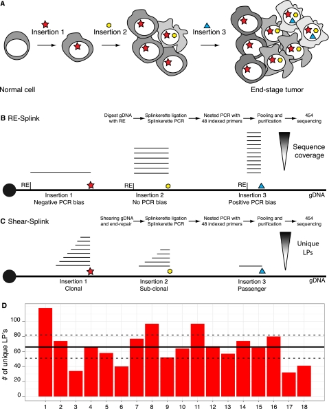
Retroviral and transposon-based insertional mutagenesis (IM) screens are widely used for cancer gene discovery in mice. Exploiting the full potential of IM screens requires methods for high-throughput sequencing and mapping of transposon and retroviral insertion sites. Current protocols are based on ligation-mediated PCR amplification of junction fragments from restriction endonuclease-digested genomic DNA, resulting in amplification biases due to uneven genomic distribution of restriction enzyme recognition sites. Consequently, sequence coverage cannot be used to assess the clonality of individual insertions. We have developed a novel method, called shear-splink, for the semiquantitative high-throughput analysis of insertional mutations. Shear-splink employs random fragmentation of genomic DNA, which reduces unwanted amplification biases. Additionally, shear-splink enables us to assess clonality of individual insertions by determining the number of unique ligation points (LPs) between the adapter and genomic DNA. This parameter serves as a semiquantitative measure of the relative clonality of individual insertions within heterogeneous tumors. Mixing experiments with clonal cell lines derived from mouse mammary tumor virus (MMTV)-induced tumors showed that shear-splink enables the semiquantitative assessment of the clonality of MMTV insertions. Further, shear-splink analysis of 16 MMTV- and 127 Sleeping Beauty (SB)-induced tumors showed enrichment for cancer-relevant insertions by exclusion of irrelevant background insertions marked by single LPs, thereby facilitating the discovery of candidate cancer genes. To fully exploit the use of the shear-splink method, we set up the Insertional Mutagenesis Database (iMDB), offering a publicly available web-based application to analyze both retroviral- and transposon-based insertional mutagenesis data.


Figures

Figure 1.
Figure 1.
Schematic overview showing the clonality of insertional mutations in tumorigenesis screens, and methods to identify the insertion sites. (A) Clonal expansion of a cell containing an insertion giving a certain growth advantage, which initiates tumorigenesis. In time, additional insertions occur, resulting in a heterogeneous tumor containing a complex collection of insertional mutations. (B) Overview of restriction enzyme based LM-PCR (RE-splink). Amplification of insertion sites using restriction enzymes results in amplicons with a fixed size introducing amplification and sequencing biases, thereby hampering a quantitative identification of insertional mutations within a tumor. (C) Overview of shearing-based LM-PCR (shear-splink), which reduces amplification and sequencing biases and allows the identification of unique ligation points of the splinkerette adapter, each representing a cell within the tumor. (D) Numbers of unique LPs identified for 18 piggyBac insertions in a clonal cell line. The average and 95% confidence interval are indicated by a solid line and dashed line, respectively.
Figure 2.
Figure 2.
Shear-splink allows semiquantitative analysis of insertional mutations. (A) Southern blot analysis with an MMTV-LTR specific probe shows (in addition to the internal MMTV fragment) three bands representing endogenous MMTV copies in the FVB genome. Clonal MMTV mammary tumor cell lines BB12 and AE6 contain different patterns of somatic MMTV insertions. (B) Overview of total sequence read numbers containing a unique LP for each mixing ratio of BB12 and AE6 DNA. (C) Mixing of BB12 and AE6 DNA does not affect the number of unique LPs for a somatic MMTV insertion that is present in both cell lines. (D,E) Somatic MMTV insertions that are unique for BB12 or AE6 show numbers of unique LPs that correlate with relative clonality of the insertions (R2 > 0.86 for five out of six insertions).
Figure 3.
Figure 3.
Analysis of insertions in MMTV-induced mammary tumors by shear-splink and RE-splink. (A) Venn diagram showing strong overlap between MMTV insertions at known CISs for the shear-splink and RE-splink methods. The overlapping CISs are strongly enriched for components of the Wnt and Fgf signaling pathways, which are known to cooperate during mammary tumorigenesis. (B) Total number of unique insertions (>0 and >1 sequence coverage and unique LPs) identified in a panel of 16 MMTV-induced tumors using shear-splink or RE-splink with BfaI or NlaIII. A high level of variability is observed in the absolute number of insertions not linked to a CIS, in contrast to a comparable number of insertions mapping to known CISs. (C) Bar diagrams showing percentages of insertions representing known CISs for shear-splink and for RE-splink with BfaI and NlaIII. Increasing the threshold to higher sequence coverage of unique LPs increases the fraction of insertions representing known CISs. For all thresholds tested (>0, >1, >2, >5, >10), the percentage of insertions mapping to known CISs is higher for shear-splink than for RE-splink. (D) Receiver Operating Characteristic (ROC) curves for the RE-splink and shear-splink methods show for shear-splink that enrichment in the identification of relevant insertions does not result in reduced sensitivity. The ROC curves are built upon unique LPs for the shear-splink analysis and sequence coverage for the RE-splink experiments. By moving along the ROC curves from left to right, the ratios between true positives (sensitivity) and false positives (specificity) are visualized.
Figure 4.
Figure 4.
Analysis of insertions in Sleeping Beauty–induced lymphomas by shear-splink and RE-splink. (A) Venn diagram showing the overlap of CISs in 127 Sleeping Beauty–induced lymphomas, as identified by shear-splink and RE-splink using BfaI or NlaIII . In total, 53 CISs are identified. Shear-splink detected more CISs (45) than BfaI- and NlaIII-based RE-splink (31 and 33 CISs, respectively). (B) Shear-splink enriches for insertions contributing to CISs, as shown for the number of CISs identified per 1000 insertions. Combining both RE-splink data sets does not increase the efficiency, since the number of CISs per 1000 insertions is similar to the individual RE-splink analysis. (C) The percentage of insertions contributing to a CIS is higher for shear-splink than for the individual or combined RE-splink datasets, confirming that shear-splink enriches for relevant insertions. (D) Insertions near cancer-related genes are represented by higher numbers of unique LPs. Density plot showing the distribution of unique LPs for SB insertions neighboring a Cancer Genome Census (CGC) gene (red) vs. those not flanking a CGC gene (gray).

References

    1. Amsterdam A, Burgess S, Golling G, Chen W, Sun Z, Townsend K, Farrington S, Haldi M, Hopkins N 1999. A large-scale insertional mutagenesis screen in zebrafish. Genes Dev 13: 2713–2724 - PMC - PubMed
    1. Bouwman P, Aly A, Escandell JM, Pieterse M, Bartkova J, van der Gulden H, Hiddingh S, Thanasoula M, Kulkarni A, Yang Q, et al. 2010. 53BP1 loss rescues BRCA1 deficiency and is associated with triple-negative and BRCA-mutated breast cancers. Nat Struct Mol Biol 17: 688–695 - PMC - PubMed
    1. Carette JE, Guimaraes CP, Varadarajan M, Park AS, Wuethrich I, Godarova A, Kotecki M, Cochran BH, Spooner E, Ploegh HL, et al. 2009. Haploid genetic screens in human cells identify host factors used by pathogens. Science 326: 1231–1235 - PubMed
    1. Collier LS, Carlson CM, Ravimohan S, Dupuy AJ, Largaespada DA 2005. Cancer gene discovery in solid tumors using transposon-based somatic mutagenesis in the mouse. Nature 436: 272–276 - PubMed
    1. Copeland NG, Jenkins NA 2010. Harnessing transposons for cancer gene discovery. Nat Rev Cancer 10: 696–706 - PubMed

Publication types

Associated data