. 2011 Oct;138(19):4267-77.
doi: 10.1242/dev.067900. Epub 2011 Aug 18.

Cell cycle-regulated multi-site phosphorylation of Neurogenin 2 coordinates cell cycling with differentiation during neurogenesis

Affiliations

Cell cycle-regulated multi-site phosphorylation of Neurogenin 2 coordinates cell cycling with differentiation during neurogenesis

Fahad Ali et al. Development. 2011 Oct.

Abstract

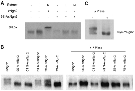
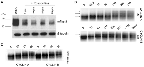
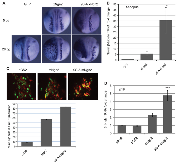
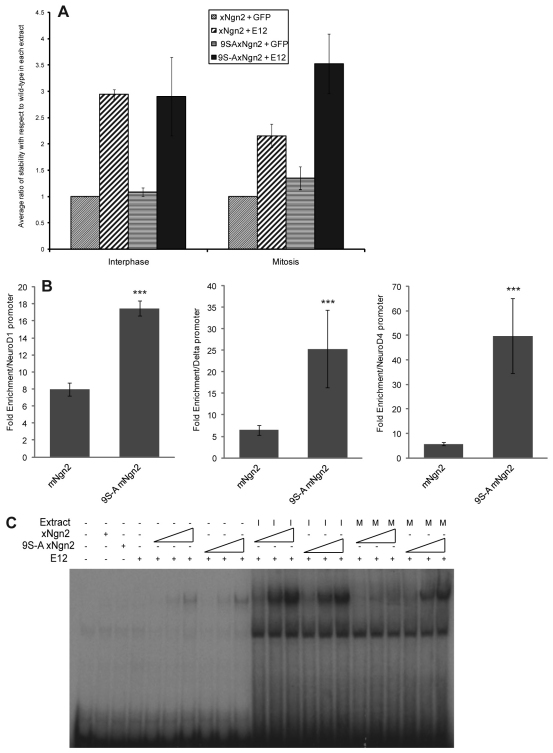
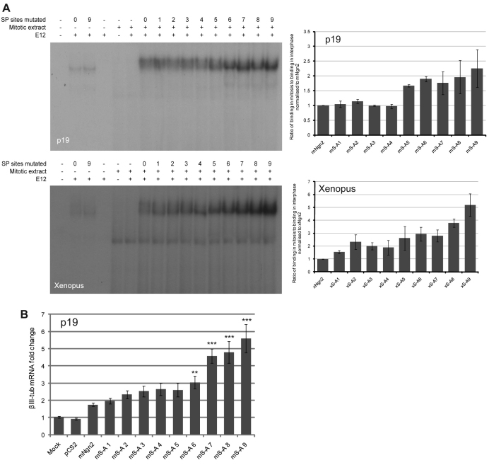
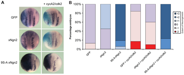
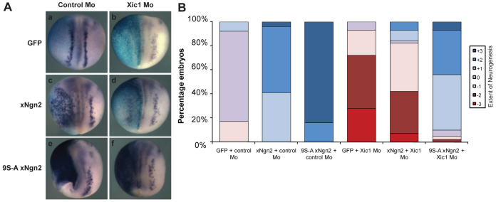
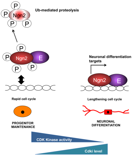
During development of the central nervous system, the transition from progenitor maintenance to differentiation is directly triggered by a lengthening of the cell cycle that occurs as development progresses. However, the mechanistic basis of this regulation is unknown. The proneural transcription factor Neurogenin 2 (Ngn2) acts as a master regulator of neuronal differentiation. Here, we demonstrate that Ngn2 is phosphorylated on multiple serine-proline sites in response to rising cyclin-dependent kinase (cdk) levels. This multi-site phosphorylation results in quantitative inhibition of the ability of Ngn2 to induce neurogenesis in vivo and in vitro. Mechanistically, multi-site phosphorylation inhibits binding of Ngn2 to E box DNA, and inhibition of DNA binding depends on the number of phosphorylation sites available, quantitatively controlling promoter occupancy in a rheostat-like manner. Neuronal differentiation driven by a mutant of Ngn2 that cannot be phosphorylated by cdks is no longer inhibited by elevated cdk kinase levels. Additionally, phosphomutant Ngn2-driven neuronal differentiation shows a reduced requirement for the presence of cdk inhibitors. From these results, we propose a model whereby multi-site cdk-dependent phosphorylation of Ngn2 interprets cdk levels to control neuronal differentiation in response to cell cycle lengthening during development.


Figures

Fig. 1.
Fig. 1.
Ngn2 undergoes cell cycle-regulated phosphorylation. (A) In vitro translated (IVT) [35S]methionine-labelled xNgn2 and 9S-A xNgn2 were incubated in interphase (I) and mitotic (M) Xenopus egg extracts before SDS-PAGE and autoradiography. In M extracts, phosphorylated xNgn2 runs at the level of the 36 kDa molecular weight marker (indicated). (B) Western blot analysis of HA-tagged mNgn2 and mutants thereof, transfected into mouse P19 cells with and without λ-phosphatase treatment. (C) Western blot analysis of myc-mNgn2 from E16.5 mouse cortex, with and without λ-phosphatase treatment.
Fig. 2.
Fig. 2.
Ngn2 is phosphorylated by cyclin-dependent kinases. (A) Western blot analysis of HA-tagged mNgn2 and 9S-A mNgn2 24 hours after transfection into mouse P19 cells with and without treatment with the cdk inhibitor roscovitine. β-Tubulin provides a loading control. (B) SDS-PAGE analysis of IVT mNgn2 in human HeLa cell extract in response to increasing doses of recombinant cyclin A (top) or cyclin B (bottom). Arrows indicate different phosphoforms of mNgn2. (C) SDS-PAGE analysis of the kinetics of phosphorylation of IVT mNgn2 when added to HeLa extracts containing recombinant cyclin A or cyclin B proteins.
Fig. 3.
Fig. 3.
Mutation of phosphorylation sites promotes Ngn2 activity. (A) Xenopus embryos were injected (left side) in one of two cells with either 5 or 20 pg mRNA as indicated, fixed at stage 15 and subject to in situ hybridisation for neural β-tubulin. The number of embryos scored was 39-90 per condition. (B) qPCR analysis of neural β-tubulin in stage 15 Xenopus embryos injected at the one-cell stage with 20 pg xNgn2 or 9S-A xNgn2 mRNA. Average fold increase in neural β-tubulin mRNA expression is shown normalised to GFP-injected control (mean ± s.e.m.; *, P≤0.05). (C) Mouse P19 cells transfected with mNgn2 or 9S-A mNgn2 with GFP were fixed 24 hours after transfection and stained for expression of neuron-specific βIII-tubulin (TuJ1) (red), quantitating TuJ1+ among GFP+ cells (mean ± s.e.m.). (D) qPCR analysis of βIII-tubulin in P19 cells 24 hours following transfection with mNgn2 and 9S-A mNgn2. Average fold increase in mRNA expression is shown normalised to housekeeping gene expression (mean ± s.e.m.; ***, P≤0.005).
Fig. 4.
Fig. 4.
Phosphomutant Ngn2 binds more efficiently than wild-type Ngn2 protein to DNA and is stabilised more by E12. (A) IVT [35S]methionine-labelled xNgn2 or 9S-A xNgn2 were incubated in I or M Xenopus egg extracts in the presence of unlabelled IVT GFP or E12. Samples were removed every 10 minutes, separated by SDS-PAGE and the amount of Ngn2 protein determined by phosphorimaging, calculating the half-life of Ngn2 protein degradation using first-order rate kinetics. Half-lives were normalised to that of xNgn2 with GFP in each experiment and the average ratios of stability relative to xNgn2 with GFP for three experiments were plotted (mean ± s.e.m.). (B) Chromatin immunoprecipitation (ChIP) analysis of cell extracts containing normalised amounts of HA-tagged mNgn2 and 9S-A mNgn2, assessing binding to the promoters of Neurod1, delta-like 1 (Dll1) and Neurod4 in mouse P19 cells, 24 hours following transfection (IgG control for non-specific background binding). ***, P≤0.005. (C) Electrophoretic mobility shift assay (EMSA) showing E box binding of normalised IVT xNgn2 or 9S-A xNgn2 in I and M extracts with and without E12.
Fig. 5.
Fig. 5.
Phosphorylation on multiple serine-proline (SP) sites has an additive effect on Ngn2 DNA binding. (A) EMSA analyses in Xenopus M extracts showing E box binding of normalised IVTs of xNgn2 or mNgn2 and additive phosphorylation site mutants of xNgn2 or mNgn2 as labelled. Graphs show quantitation of E box binding of phosphorylation site mutants by phosphorimaging, normalised to DNA binding of xNgn2 or mNgn2 as appropriate (see Fig. S1 in the supplementary material). (B) qPCR analysis of neural βIII-tubulin in P19 cells. P19 cells were transiently transfected with mNgn2 and additive phosphorylation site mutants thereof for 24 hours. Average fold increase (± s.e.m.) in βIII-tubulin mRNA expression normalised to housekeeping genes [β-actin (Actb) and Gapdh]. **, P≤0.05; ***, P≤0.005.
Fig. 6.
Fig. 6.
9S-A xNgn2 is resistant to cyclin A2/cdk2-mediated suppression of neurogenesis in vivo. (A) Xenopus embryos were injected (left side) in one of two cells with 20 pg xNgn2 or 9S-A xNgn2 mRNA, together with 500 pg cyclin A2 and cdk2 mRNA, assaying for expression of neural β-tubulin at stage 15 by in situ hybridisation. (B) Semi-quantitative analysis of in situ hybridisation data. The number of embryos scored was 48-86 per condition. Neurogenesis was enhanced (+3, +2, +1), the same as (0) or reduced (–1, –2, –3) compared with the uninjected side (see Fig. S8 in the supplementary material).
Fig. 7.
Fig. 7.
9S-A xNgn2 does not require the cdk inhibitor Xic1 for activity. (A) Xenopus embryos were injected (left side) in one of four cells (dorsally targeted) with 20 pg mRNA as indicated, together with 20 ng of either control (a,c,e) or Xic1 (b,d,f) morpholino, fixed at stage 15 and subject to in situ hybridisation for neural β-tubulin expression. (B) Semi-quantitative analysis of in situ hybridisation data. The number of embryos scored was 43-59 per condition. Neurogenesis was enhanced (+3, +2, +1), the same as (0) or reduced (–1, –2, –3) compared with the uninjected side (see Fig. S8 in the supplementary material for examples of the scoring method).
Fig. 8.
Fig. 8.
Model of control of neuronal differentiation via cell cycle length and Ngn2 phosphorylation. Phosphorylation of Ngn2 protein occurs in rapid progenitor cell cycles. Cell cycle lengthening results in an accumulation of un(der)phosphorylated Ngn2, enhancing promoter binding and resulting in the activation of downstream target genes that drive differentiation. E, E protein; Ub, ubiquitin; P, phosphorylation.

Similar articles

Cited by

References

    1. Aguado-Llera D., Goormaghtigh E., de Geest N., Quan X. J., Prieto A., Hassan B. A., Gomez J., Neira J. L. (2010). The basic helix-loop-helix region of human neurogenin 1 is a monomeric natively unfolded protein which forms a ``fuzzy'' complex upon DNA binding. Biochemistry 49, 1577-1589 - PubMed
    1. Bain J., Plater L., Elliott M., Shpiro N., Hastie C. J., McLauchlan H., Klevernic I., Arthur J. S., Alessi D. R., Cohen P. (2007). The selectivity of protein kinase inhibitors: a further update. Biochem. J. 408, 297-315 - PMC - PubMed
    1. Bertrand N., Castro D. S., Guillemot F. (2002). Proneural genes and the specification of neural cell types. Nat. Rev. Neurosci. 3, 517-530 - PubMed
    1. Crane-Robinson C., Dragan A. I., Privalov P. L. (2006). The extended arms of DNA-binding domains: a tale of tails. Trends Biochem. Sci. 31, 547-552 - PubMed
    1. Cremisi F., Philpott A., Ohnuma S. (2003). Cell cycle and cell fate interactions in neural development. Curr. Opin. Neurobiol. 13, 26-33 - PubMed

Publication types

Substances