. 2011 Oct;138(19):4245-54.
doi: 10.1242/dev.070433. Epub 2011 Aug 18.

Notch pathway activation can replace the requirement for Wnt4 and Wnt9b in mesenchymal-to-epithelial transition of nephron stem cells

Affiliations

Notch pathway activation can replace the requirement for Wnt4 and Wnt9b in mesenchymal-to-epithelial transition of nephron stem cells

Scott C Boyle et al. Development. 2011 Oct.

Abstract

The primary excretory organ in vertebrates is the kidney, which is responsible for blood filtration, solute homeostasis and pH balance. These functions are carried out by specialized epithelial cells organized into tubules called nephrons. Each of these cell types arise during embryonic development from a mesenchymal stem cell pool through a process of mesenchymal-to-epithelial transition (MET) that requires sequential action of specific Wnt signals. Induction by Wnt9b directs cells to exit the stem cell niche and express Wnt4, which is both necessary and sufficient for the formation of epithelia. Without either factor, MET fails, nephrons do not form and newborn mice die owing to kidney failure. Ectopic Notch activation in stem cells induces mass differentiation and exhaustion of the stem cell pool. To investigate whether this reflected an interaction between Notch and Wnt, we employed a novel gene manipulation strategy in cultured embryonic kidneys. We show that Notch activation is capable of inducing MET in the absence of both Wnt4 and Wnt9b. Following MET, the presence of Notch directs cells primarily to the proximal tubule fate. Only nephron stem cells have the ability to undergo MET in response to Wnt or Notch, as activation in the closely related stromal mesenchyme has no inductive effect. These data demonstrate that stem cells for renal epithelia are uniquely poised to undergo MET, and that Notch activation can replace key inductive Wnt signals in this process. After MET, Notch provides an instructive signal directing cells towards the proximal tubule lineage at the expense of other renal epithelial fates.


Figures
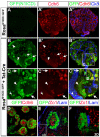
Fig. 1.
Fig. 1.
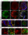
Tat-Cre mediates efficient recombination in cultured kidneys and a subset of cells undergo MET in response to N1ICD. (A-B′) E12.5 RosaN1ICD::GFP kidneys cultured with vehicle (A-A′) or 1 γM Tat-Cre (B-B′) and grown for 72 hours. GFP (green) marks cells that have undergone recombination: Cdh6 (red) indicates PT differentiation and Ck8 (blue) labels the collecting system. GFP+ cells are only detected in Tat-Cre treated kidneys. (C-C′) High-power image of area boxed in B′. A subset of GFP+ cells forms Cdh6+ clusters in response to N1ICD (arrows in B-C′), whereas others do not (arrowheads in B-C′). The collecting duct is largely resistant to Tat-Cre delivery, but Ck8+, GFP+, Cdh6+ cells were never observed (C-C′, concave arrowheads). (D) High-power image of area boxed in C′ demonstrating morphology GFP+, Cdh6+ clusters. (E,F) Single plane (E) and 3D-reconstruction (F) images indicate presence of Zo1-expressing tight junctions (red) and deposition of a stratified laminin basement membrane (blue), demonstrating that GFP+ clusters are epithelial cells. Inset in E shows staining of duct epithelium in the same kidney.
Fig. 2.
Fig. 2.
Expression of N1ICD limits potential to PT and early podocyte lineages. N1ICD clusters were stained with markers of differentiated nephron segments. (A,B) Examples of Aqp1- (A) and LTL- (B) positive cells indicate fully differentiated PT. (C) Clusters consistently co-expressed Aqp1 and LTL, indicative of PT, but not the loop of Henle (Aqp1+,LTL). (D) Wt1high clusters were found at much lower frequency, and indicate presence of immature podocytes. No glomerular organization was observed in these clusters. (E-H) GFP+ cells did not express markers of more distal nephron segments, including thick ascending limb (E, Tamm Horsfall protein), distal tubule (F, E-cadherin; G, Clcknb) or connecting segment (H, Cd28K). Insets in E,F,H demonstrate the fidelity of antibodies in the same explants. Scale bars: 50 γm. Arrows indicate low levels of E-cadherin in N1ICD::GFP-expressing clusters; arrowheads indicate level and localization normally seen in duct.
Fig. 3.
Fig. 3.
Only cap mesenchyme undergoes MET in response to N1ICD. N1ICD was expressed in vivo in cap (Six2Cre) and stromal (FoxD1Cre) mesenchyme to assess response. (A-C) Expression of CMM marker Cited1 (A), PT marker Cdh6 (B) and CMM/early podocyte marker Wt1 (C) in wild-type kidneys. (D) Expression of N1ICD in the CMM exhausts the Cited1+ stem cell pool. (E,F) CMM responding to N1ICD adopt the PT (Cdh6, E) and early podocyte (Wt1high, F) fates. (G) Cited1-expressing stem cells (red) are maintained when N1ICD is expressed in the stromal mesenchyme. (H) The vast majority of stromal cells do not undergo MET in response to N1ICD. Small Cdh6+ clusters are rarely detected in the stroma (inset). (I) N1ICD+ stromal cells do not express Wt1. Scale bars: 100 γm.
Fig. 4.
Fig. 4.
Proximal differentiation defects in Notch mutants are not due to loss of Wnt4. (A,B) In situ hybridization for Wnt4 mRNA in E15.5 control (A, Pax3tgCre; N2+/f) and Notch loss-of-function kidneys (B, Pax3tgCre; N2f/f). Wnt4 expression is maintained in the absence of Notch2. (C-F) Control E12.5 explants grown on either 3T3-lacZ (C,D) or 3T3-Wnt4 cells (E,F) readily form LTL+ PT (C,E, green) and synaptopodin+ (D,F, green), Wt1+ (D,F, red) glomeruli. (G-J) Exogenous Wnt4 does not rescue proximal fates in Notch mutants. PT (G,I) and glomeruli (H,J) are absent in Pax3Cre,Notch2f/f explants, demonstrated by absence of LTL and synaptopodin/glomerular Wt1 in kidneys cultured on control (G,H) and Wnt4-expressing cells (I,J).
Fig. 5.
Fig. 5.
Wnt4 is not required for MET in response to N1ICD. (A-A′) CMM in Wnt4–/– kidneys does not undergo MET. An occasional Cdh6+ cluster can be detected (A′, inset), but no functional nephrons are made. (B-B′) N1ICD rescues MET in Wnt4–/– kidneys. Numerous GFP+/Cdh6+ clusters are found near branched UB tips (arrows). Like wild type, the stroma does not respond (arrowheads). (C) High-power image of boxed area in B′.(D-D′) N1ICD clusters in Wnt4–/– explants are epithelia; they form Zo1+ tight junctions (red) and deposit basement membrane (blue). (E-F′) N1ICD cells in Wnt4–/– explants progress to Aqp1+ PT epithelia (E) and Wt1high immature podocytes (F). Scale bars: 50 γm.
Fig. 6.
Fig. 6.
N1ICD can induce MET independently of Wnt9b. (A-A′) No epithelia are formed in control Wnt9b–/– kidneys. (B-B′) MET is restored in Tat-Cre treated RosaN1ICD, Wnt9b–/– kidneys, as demonstrated by numerous GFP+/Cdh6+ clusters around UB tips. (C,C′) High-magnification image of boxed area in B′. (D,D′) N1ICD cells in Wnt9b–/– explants are epithelia; they form Zo1+ tight junctions (red) and deposit basement membrane (blue). (E-F′) N1ICD cells in Wnt9b–/– make Aqp1+ PT epithelia (E) and Wt1high immature podocytes (F). Scale bars: 50 γm. (G) Distribution of fate decisions among cells responding to N1ICD in wild-type, Wnt4–/– and Wnt9b–/– kidneys. Data are mean+s.e.m. (H) Semi-quantitative RT-PCR on kidneys from wild-type and Wnt9b–/– explants. Lane 1, wild-type control; lane 2, wild-type Tat-Cre treated; lane 3, Wnt9b–/– control; lane 4, Wnt9b–/– Tat-Cre treated (24 hours); lane 5, Wnt9b–/– Tat-Cre treated (48 hours); lane 6, wild-type Tat-Cre treated (no-RT). Markers of all transitional and mature cell types are detected in both wild-type and wild-type Tat-Cre-treated kidneys. Wnt9–/– kidneys express only markers of CMM. Wnt9b–/– kidneys treated with Tat-Cre express markers indicative of normal PT development, including Fgf8, Wnt4 (PTA), Lhx1, Brn1, Dll1 (RV), Hes1, Hes5 and Papss2 (SSB). Expression of PT markers has a temporal component, Cdh6 is detected at 24 hours, but Aqp1 not until 48 hours. The podocyte marker Greb1 is expressed, but not the mature marker podocin. Treated and untreated explants do not make distal epithelium (Clcnkb).
Fig. 7.
Fig. 7.
Wnt4/Wnt9b double mutants make epithelia in response to N1ICD. (A-A′) MET does not occur in Wnt9b–/–,Wnt4–/– kidneys. (B-B′) MET occurs in Tat-Cre treated RosaN1ICD,Wnt9b–/–,Wnt4–/– kidneys, as evidenced by many GFP+/Cdh6+ clusters around UB tips. (C,C′) High magnification image of boxed area in B′. Position of GFP+/Cdh6+ cells is consistent with MET of CMM in the absence of both Wnt9b and Wnt4. (D,D′) N1ICD cells in Wnt9b–/–,Wnt4–/– explants are epithelia; they form Zo1+ tight junctions (red) and deposit basement membrane (blue). (E-F′) N1ICD cells in double mutants can make Aqp1+ PT epithelia (E, arrows) and Wt1high immature podocytes (F, arrows). Scale bars: 50 γm.

References

    1. Barak H., Boyle S. C. (2011). Organ culture and immunostaining of mouse embryonic kidneys. Cold Spring Harb. Protoc. 2011, doi: 10.1101/pdb prot5558 - PubMed
    1. Barasch J., Yang J., Ware C. B., Taga T., Yoshida K., Erdjument-Bromage H., Tempst P., Parravicini E., Malach S., Aranoff T., et al. (1999). Mesenchymal to epithelial conversion in rat metanephros is induced by LIF. Cell 99, 377-386 - PubMed
    1. Blair S. S. (1996). Notch and Wingless signals collide. Nature 271, 1822-1823 - PubMed
    1. Boyle S., Shioda T., Perantoni A. O., de Caestecker M. (2007). Cited1 and Cited2 are differentially expressed in the developing kidney but are not required for nephrogenesis. Dev. Dyn. 236, 2321-2330 - PubMed
    1. Boyle S., Misfeldt A., Chandler K. J., Deal K. K., Southard-Smith E. M., Mortlock D. P., Baldwin H. S., de Caestecker M. (2008). Fate mapping using Cited1-CreERT2 mice demonstrates that the cap mesenchyme contains self-renewing progenitor cells and gives rise exclusively to nephronic epithelia. Dev. Biol. 313, 234-245 - PMC - PubMed

Publication types