Review
. 2011 Jul-Aug;12(4):487-98.
doi: 10.3348/kjr.2011.12.4.487. Epub 2011 Jul 22.

MRI findings of rectal submucosal tumors

Affiliations
Review

MRI findings of rectal submucosal tumors

Honsoul Kim et al. Korean J Radiol. 2011 Jul-Aug.

Abstract

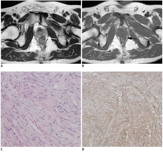
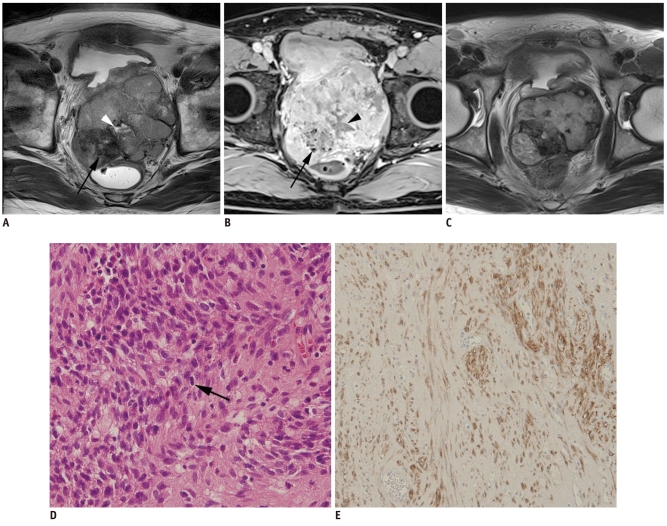
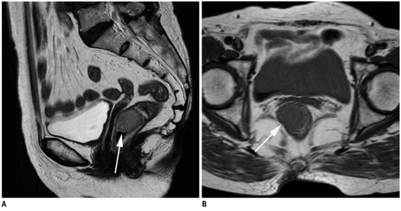
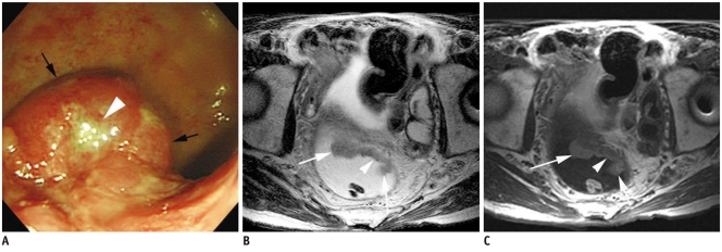
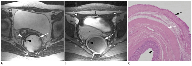
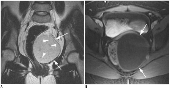
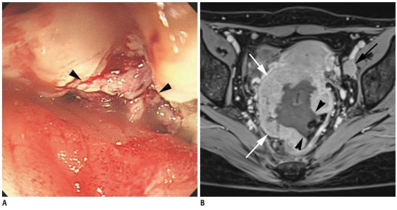
Rectal submucosal lesions encompass a wide variety of benign and malignant tumors involving the rectum. With optical colonoscopy, any mass-like protrusion covered by normal mucosa, whether the underlying process is intramural or extramural in origin, may be reported as a submucosal lesion. Whereas the assessment of submucosal lesions may be limited with performing optical colonoscopy, cross-sectional imaging such as CT, transrectal ultrasonography and MRI allows the evaluation of perirectal tissues and pelvic organs in addition to the entire thickness of the rectum, and so this is advantageous for the assessment of rectal submucosal tumors. Among these, MRI is the best investigative modality for soft tissue characterization. Therefore, knowledge of the MRI features of rectal submucosal tumors can help achieve accurate preoperative diagnoses and facilitate the appropriate management.

Keywords: MRI; Rectal cancer; Rectal submucosal tumor.


Figures

Fig. 1
Fig. 1
Rectal leiomyoma in 42-year-old woman. Axial T2-weighted image (A), T1-weighted image (B) and gadolinium enhanced axial T1-weighted image (C) demonstrated submucosal mass (arrows) in anterior wall of distal rectum. Mass is well-demarcated and it shows signal intensity that is slightly brighter than that of skeletal muscles on T2-weighted image.
Fig. 2
Fig. 2
Rectal lipoma in 74-year-old man. Axial T1-weighted image (A) demonstrates oval shaped rectal submucosal mass (arrow) with signal density identical to that of perirectal fat tissue. On gadolinium enhanced axial T1-weighted image (B) signal of lesion drops due to fat suppression, which confirms diagnosis of lipoma (arrow).
Fig. 3
Fig. 3
Rectal carcinoid (well-differentiated neuroendocrine tumor) in 60-year-old man. Axial T2-weighted image (A), axial T1-weighted image (B) and gadolinium enhanced axial T1-weighted image with fat suppression (C) show discrete 2.5-cm homogeneously enhancing intraluminal submucosal mass in rectum (arrows).
Fig. 4
Fig. 4
Poorly-differentiated neuroendocrine carcinoma in 61-year-old woman which was diagnosed as small cell carcinoma. Colonoscopy (A) shows ulcerofungating mass (white arrows) in lower rectum, which is similar to adenocarcinoma. Axial T2-weighted image (B) and gadolinium enhanced coronal T1-weighted image (C) also demonstrate fungating mass (white arrows) in lower rectum. Enlarged metastatic lymph node (arrowheads) is observed in mesorectum.
Fig. 5
Fig. 5
Rectal gastrointestinal stromal tumor in 59-year-old woman. Axial T2-weighted image (A) and T1-weighted image (B) show 1.5-cm submucosal mass (arrows) in left anterior lateral wall of lower rectum. Mass is relatively well-demarcated. After ultralow anterior resection, histopathologic diagnosis was very low-risk gastrointestinal stromal tumor (mitotic rate: 0 per 50 high power fields) based on Hematoxylin & Eosin staining (× 200) (C) and positive c-kit results on immunohistochemistry (D).
Fig. 6
Fig. 6
Rectal gastrointestinal stromal tumor in 53-year-old man. Axial T2-weighted image (A) and gadolinium enhanced axial T1-weighted image with fat suppression (B) revealed 9.6-cm multilobulated mass that was possibly arising from anterior wall of rectum. Within mass, dark signal intensities (arrows) due to internal calcification and fluid intensity (arrowheads) due to necrosis are seen. After imatinib treatment, prominent tumor shrinkage is demonstrated on follow-up axial T2-weighted image (C). Mile's operation was performed. Hematoxylin & Eosin staining (× 200) (D) and positive results of c-kit immunohistochemistry (E) revealed diagnosis of high-risk gastrointestinal stromal tumor, in which frequent mitosis (arrow in D; mitotic rate: more than 10 per 50 high power fields) was observed.
Fig. 7
Fig. 7
Rectal schwannoma in 67-year-old woman. Sagittal T2-weighted image (A) and axial T1-weighted image (B) show well-circumscribed homogeneous submucosal mass (arrows) in lower rectum.
Fig. 8
Fig. 8
Primary rectal lymphoma in 62-year-old man. Optical colonoscopy (A) identified fungating mass (black arrows) with central necrosis (white arrowhead). Axial T2-weighted image (B) and T1-weighted image (C) show 4.5-cm well circumscribed fungating mass (arrows) with central ulceration (arrowheads) located at upper rectum. Homogeneous tumoral signal intensity and preservation of fat planes may suggest lymphoma.
Fig. 9
Fig. 9
Malignant melanoma in 66-year-old woman. Sagittal T2-weighted image (A) and axial T1-weighted image (B) show bulky intraluminal fungating mass (arrows) involving distal rectum. Perirectal infiltration and necrotic nodal metastases (arrowhead) in posterior side of mid rectum are seen. Signal intensity of this mass is heterogeneously high on T2-weighted image and low on T1-weighted image. After performing Mile's operation, amelanocytic melanoma with lymph node metastases was confirmed.
Fig. 10
Fig. 10
Endometriosis that presented as rectal submucosal mass in 46-year-old woman. Sagittal T2-weighed image (A) shows areas of irregular tissue signal intensity in rectouterine pouch with internal cyst-like structure (arrowhead). Infiltrative rectal wall thickening (arrows) suggests rectal wall involvement. This cyst was identified as hemorrhagic structure by high signal intensity after fat suppression on axial T1-weighted image (arrowhead) (B). Gadolinium enhanced axial T1-weighted image with fat suppression (C) demonstrates heterogeneous enhancing infiltrative mass involving anterior rectal wall and rectouterine pouch (arrows). Lower anterior resection was performed, and endometriosis involvement in submucosa, proper muscle and subserosa of rectum was diagnosed.
Fig. 11
Fig. 11
Tailgut cyst in 47-year-old woman. Sagittal T2-weighted image (A) shows multilocular cystic mass with variable signal intensities (arrows) posterior to rectum (arrowheads). Gadolinium-enhanced axial T1-weighted image with fat suppression (B) also shows thin peripheral cystic wall enhancement without enhancing solid portions (arrow). On sectioning (not shown), surgical specimen revealed brownish friable material and microscopic examination demonstrated mucin-containing columnar epithelium, which is consistent with tailgut cyst.
Fig. 12
Fig. 12
Adenocarcinoma arising from tailgut cyst in 40-year-old woman. Axial T2-weighted image (A) and axial T1-weighted image (B) revealed infiltrative soft tissue mass (arrowheads) involving right gluteus muscle and coccygeal area. Retrorectal multilocular cystic mass is adjacent to this infiltrative mass (arrows). Histopathologic diagnosis was poorly-differentiated adenocarcinoma arising from tailgut cyst.
Fig. 13
Fig. 13
Duplication cyst in 28-year-old woman. Axial T2-weighted image (A) and gadolinium enhanced axial T1-weighted image with fat suppression (B) show retrorectal unilocular cystic structure with relatively thick cystic wall enhancement (white arrows) that is displacing rectum (white arrowhead). There was no communication between lesion and rectal lumen on barium study (not shown). Peripheral calcifications are seen as focal dark signal intensities (black arrowheads), which were confirmed by CT. However, calcification in alimentary duplication cyst is extremely rare finding. Microscopic assessment after surgical excision (C) revealed ectopic muscle layer (white arrow) located at subserosal compartment of rectum (black arrowhead: rectal mucosa). Superficial to this muscle layer lies submucosa and mucosa (black arrow) layer of cystic lesion.
Fig. 14
Fig. 14
Epidermoid cyst in 31-year-old woman. Oblique coronal T2-weighted image (A) shows thin-walled unilocular cystic lesion (arrows) lateral to rectum. Note small linear hypointense structures (arrowheads) that possibly represent keratin aggregates. Gadolinium enhanced axial T1-weighted image with fat suppression (B) revealed mild enhancement of cystic wall (arrows) without evidence of enhancing soft tissue components.
Fig. 15
Fig. 15
Ovarian cancer mimicking rectal intramural submucosal tumor in 44-year-old woman who recently developed hematochezia. Optical colonoscopy (A) demonstrates large intramural submucosal tumor-like lesion with central ulceration (black arrowheads). Gadolinium enhanced axial T1-weighted image with fat suppression (B) shows 4-cm mass (white arrows) with large central ulceration (black arrowheads). Possibility of intramural mesenchymal tumors such as gastrointestinal stromal tumor was suggested on MRI interpretation. However, presence of enlarged lymph node in left pelvic side wall (black arrow), indicating possible nodal metastases, is inconsistent with this diagnosis since nodal metastasis is extremely rare in gastrointestinal stromal tumor cases. After operation, diagnosis was poorly-differentiated serous adenocarcinoma arising from right ovary with direct invasion into rectum.

References

    1. Pickhardt PJ, Kim DH, Menias CO, Gopal DV, Arluk GM, Heise CP. Evaluation of submucosal lesions of the large intestine: part 1. Neoplasms. Radiographics. 2007;27:1681–1692. - PubMed
    1. Pickhardt PJ, Kim DH, Menias CO, Gopal DV, Arluk GM, Heise CP. Evaluation of submucosal lesions of the large intestine: part 2. Nonneoplastic causes. Radiographics. 2007;27:1693–1703. - PubMed
    1. Rouse HC, Godoy MC, Lee WK, Phang PT, Brown CJ, Brown JA. Imaging findings of unusual anorectal and perirectal pathology: a multi-modality approach. Clin Radiol. 2008;63:1350–1360. - PubMed
    1. Kloppel G, Perren A, Heitz PU. The gastroenteropancreatic neuroendocrine cell system and its tumors: the WHO classification. Ann N Y Acad Sci. 2004;1014:13–27. - PubMed
    1. Okamoto Y, Fujii M, Tateiwa S, Sakai T, Ochi F, Sugano M, et al. Treatment of multiple rectal carcinoids by endoscopic mucosal resection using a device for esophageal variceal ligation. Endoscopy. 2004;36:469–470. - PubMed