. 2011 Aug;7(8):e1002186.
doi: 10.1371/journal.ppat.1002186. Epub 2011 Aug 11.

Differential contribution of PB1-F2 to the virulence of highly pathogenic H5N1 influenza A virus in mammalian and avian species

Affiliations

Differential contribution of PB1-F2 to the virulence of highly pathogenic H5N1 influenza A virus in mammalian and avian species

Mirco Schmolke et al. PLoS Pathog. 2011 Aug.

Abstract

Highly pathogenic avian influenza A viruses (HPAIV) of the H5N1 subtype occasionally transmit from birds to humans and can cause severe systemic infections in both hosts. PB1-F2 is an alternative translation product of the viral PB1 segment that was initially characterized as a pro-apoptotic mitochondrial viral pathogenicity factor. A full-length PB1-F2 has been present in all human influenza pandemic virus isolates of the 20(th) century, but appears to be lost evolutionarily over time as the new virus establishes itself and circulates in the human host. In contrast, the open reading frame (ORF) for PB1-F2 is exceptionally well-conserved in avian influenza virus isolates. Here we perform a comparative study to show for the first time that PB1-F2 is a pathogenicity determinant for HPAIV (A/Viet Nam/1203/2004, VN1203 (H5N1)) in both mammals and birds. In a mammalian host, the rare N66S polymorphism in PB1-F2 that was previously described to be associated with high lethality of the 1918 influenza A virus showed increased replication and virulence of a recombinant VN1203 H5N1 virus, while deletion of the entire PB1-F2 ORF had negligible effects. Interestingly, the N66S substituted virus efficiently invades the CNS and replicates in the brain of Mx+/+ mice. In ducks deletion of PB1-F2 clearly resulted in delayed onset of clinical symptoms and systemic spreading of virus, while variations at position 66 played only a minor role in pathogenesis. These data implicate PB1-F2 as an important pathogenicity factor in ducks independent of sequence variations at position 66. Our data could explain why PB1-F2 is conserved in avian influenza virus isolates and only impacts pathogenicity in mammals when containing certain amino acid motifs such as the rare N66S polymorphism.


Conflict of interest statement

The authors have declared that no competing interests exist.

Figures
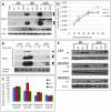
Figure 1
Figure 1. Effects of PB1-F2 expression on replication of A/Viet Nam/1203/2004 in murine cells in vitro.
A) Time course of viral protein expression. Murine lung epithelial cells (LA-4) were infected with 2MOI of A/Viet Nam/1203/2004 wild type, dF2 or N66S. Cells were lysed after indicated time points. Whole cell extracts were analyzed by western blot with specific antibodies against PB1-F2 (upper two panels, short and long exposure, respectively, PB1-F2 is indicated by black arrows), viral nucleoprotein (NP) or beta-Actin. B) PB1-F2 expression in presence or absence of 10 µM lactocystin. LA4 were infected with 2MOI of A/Viet Nam/1203/2004 wild type, dF2 or N66S. Cells were lysed after 24 h. Whole cell extracts were analyzed by western blot with specific antibodies against PB1-F2 (upper three panels, short, medium and long exposure, respectively, PB1-F2 is indicated by black arrows), equal loading is indicated by an unspecific background band (lower panel). C) Polymerase Assay. Murine fibroblasts (3T3) were transfected with indicated amounts of pCAGGS-PB1 A/Viet Nam/1203/2004 wild type (blue), dF2 (red) or N66S (green) and constant amounts of PB2, PA, NP and reporter gene expressing plasmids as described in Material and Methods. Mean firefly-luciferase activity of three independent transfections normalized to renilla-luciferase activity is depicted. Error bars represent SD. No significant differences were detected. D) Multi-cycle growth curve of A/Viet Nam/1203/2004 wild type (blue), dF2 (red) or N66S (green) on LA-4. Cells were infected with 0.05MOI of virus. Viral supernatants were taken at the indicated time points. Viral titers were determined by standard plaque assay on MDCK in absence of trypsin. Mean pfu/ml of independent triplicates ±SD indicated by the error bar are depicted. P-values are indicated (*p<0.05) using WT as standard. E) Time course of viral protein expression in LA-4 (upper panel), bone marrow derived macrophages (BMDM, middle panel) and bone marrow derived dendritic cells (BMDDC). Cells were infected with 2MOI of A/Viet Nam/1203/2004 wild type, dF2 or N66S and lysed after indicated time points. Whole cell extracts were analyzed by western blot with specific antibodies against viral nucleoprotein (NP) and tubulin.
Figure 2
Figure 2. PB1-F2 alters host response against A/Viet Nam/1203/2004 in murine cells.
A–C) LA-4 (A), BMDM (B) and BMDDC (C) were infected with 2 MOI of A/Viet Nam/1203/2004 wild type (blue), dF2 (red) or N66S (green). Transcriptional response was measured by gene specific analysis of cDNA amounts from total lysates 8 h post infection. Expression is depicted as mean nfold of mock samples +SD from three independent samples. P-values are indicated (*p<0.05, ** p<0.01) using WT as standard.
Figure 3
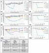
Figure 3. Effects of PB1-F2 expression on viral pathogenesis in vivo.
A–C) Weight loss curves of C57/BL/6/A2G-Mx1 mice infected with indicated doses of A/Viet Nam/1203/2004 wild type (A), dF2 (B) or N66S (C). Animals were excluded from analysis when reaching less than 75% initial body weight. Mean %-body weight of 5-9 (initial group size) animals normalized to initial weight +SD is depicted. D–F) Survival curves of C57/BL/6/A2G-Mx1 mice infected with indicated doses of A/Viet Nam/1203/2004 wild type (D), dF2 (E) or N66S (F). %-survival of 5–9 (initial group size) animals is depicted. (G) MLD50 of A/Viet Nam/1203/2004 wild type, dF2 or N66S (high and low pathogenic version) in C57/BL/6/A2G-Mx1 and C57/BL/6 mice.
Figure 4
Figure 4. Effects of PB1-F2 expression on viral replication and spreading to the brain in mice.
A) C57/BL/6/A2G-Mx1 mice were infected with 2.5×10E6 pfu of VN1203 wild type (blue), dF2 (red) or N66S (green). Viral lung titers were determined on day 2 and 5 post infection in 3 animals by standard plaque assay on MDCK cells. Titers of individual animals (pfu/ml) are depicted, the detect ion limit of the assay was 50 pfu/ml. P-values for the mean plaque titers are indicated (*p<0.05) using WT as standard. B) C57/BL/6/A2G-Mx1 mice were infected with 2.5×10E3 pfu of VN1203 wild type (blue), dF2 (red) or N66S (green). Viral brain titers on day 8 post infection of individual animals are depicted. The detection limit of the assay was 50 pfu/ml. C) Direct brain-inoculation of VN1203 wild type (blue), dF2 (red) or N66S (green) (10pfu upper panel, 100pfu lower panel). Viral brain titers (pfu/ml) 48 h post infection are shown for each animal.
Figure 5
Figure 5. Effects of PB1-F2 expression on replication of A/Viet Nam/1203/2004 in duck fibroblasts in vitro.
A) Time course of viral protein expression. Duck embryonic fibroblasts (DEF) were infected with 2MOI of A/Viet Nam/1203/2004 wild type, dF2 or N66S. Cells were lysed after indicated time points. Whole cell extracts were analyzed by western blot with specific antibodies against PB1-F2 (upper two panels, short and long exposure, respectively), viral nucleoprotein (NP) or beta-Actin. B) PB1-F2 expression in presence or absence of 10 µM lactocystin. DEF were infected with 2MOI of A/Viet Nam/1203/2004 wild type, dF2 or N66S. Cells were lysed after 24 h. Whole cell extracts were analyzed by western blot with specific antibodies against PB1-F2 (upper three panels, short, medium and long exposure, respectively, PB1-F2 is indicated by black arrows), equal loading is indicated by GAPDH levels (lower panel). C) Multi-cycle growth curve of A/Viet Nam/1203/2004 wild type (blue), dF2 (red) or N66S (green) on DEF. Cells were infected with 0.05MOI of virus. Viral supernatants were taken at the indicated time points. Viral titers were determined by standard plaque assay on MDCK in absence of trypsin. Mean pfu/ml of independent triplicates ±SD indicated by the error bar are depicted.
Figure 6
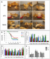
Figure 6. Diminished pathogenicity of PB1-F2 deficient virus in ducks.
Ten 2-week old white Peking ducks were infected with 10E4 pfu of A/Viet Nam/1203/2004 wild type, dF2 or N66S. A) Three representative pictures of infected ducks were taken on day 4 p.i.. B) Survival curves of 10 2-week old white peking ducks were infected with 10E4 pfu of A/Viet Nam/1203/2004 wild type (blue), dF2 (red) or N66S (green). Survival (%) is depicted. C and D) Viral titers of indicated organs from day 1 (C) and day 3 (D) post infection with wild type (blue), dF2 (red) or N66S (green). TCID50/ml of organs from individual animals are depicted. E) Mean viral titers (TCID50/ml) cloacal swabs from 10 animals per group (initial group size) are depicted. P-values are indicated (*p<0.05, ** p<0.01) using dF2 as standard.

Similar articles

Cited by

References

    1. No authors listed. Isolation of avian influenza A(H5N1) viruses from humans—Hong Kong, May-December 1997. MMWR Morb Mortal Wkly Rep. 1997;46:1204–1207. - PubMed
    1. Shinya K, Ebina M, Yamada S, Ono M, Kasai N, et al. Avian flu: influenza virus receptors in the human airway. Nature. 2006;440:435–436. - PubMed
    1. Yuen KY, Chan PK, Peiris M, Tsang DN, Que TL, et al. Clinical features and rapid viral diagnosis of human disease associated with avian influenza A H5N1 virus. Lancet. 1998;351:467–471. - PubMed
    1. Tran TH, Nguyen TL, Nguyen TD, Luong TS, Pham PM, et al. Avian influenza A (H5N1) in 10 patients in Vietnam. N Engl J Med. 2004;350:1179–1188. - PubMed
    1. Beigel JH, Farrar J, Han AM, Hayden FG, Hyer R, et al. Avian influenza A (H5N1) infection in humans. N Engl J Med. 2005;353:1374–1385. - PubMed

Publication types

Substances