. 2011 Aug;7(8):e1002225.
doi: 10.1371/journal.pgen.1002225. Epub 2011 Aug 11.

Suppression of scant identifies Endos as a substrate of greatwall kinase and a negative regulator of protein phosphatase 2A in mitosis

Affiliations

Suppression of scant identifies Endos as a substrate of greatwall kinase and a negative regulator of protein phosphatase 2A in mitosis

Hélène Rangone et al. PLoS Genet. 2011 Aug.

Abstract

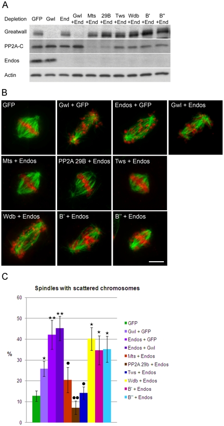
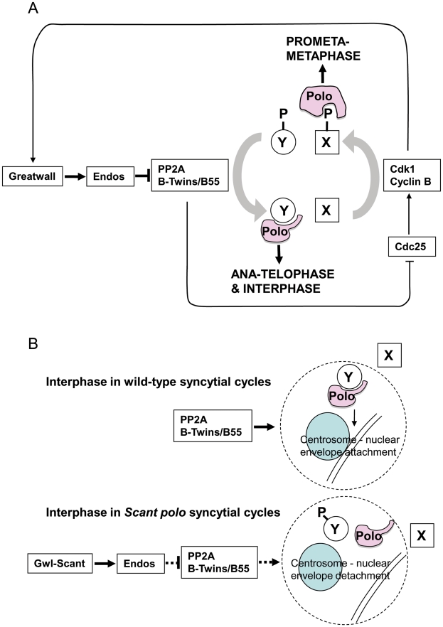
Protein phosphatase 2A (PP2A) plays a major role in dephosphorylating the targets of the major mitotic kinase Cdk1 at mitotic exit, yet how it is regulated in mitotic progression is poorly understood. Here we show that mutations in either the catalytic or regulatory twins/B55 subunit of PP2A act as enhancers of gwl(Scant), a gain-of-function allele of the Greatwall kinase gene that leads to embryonic lethality in Drosophila when the maternal dosage of the mitotic kinase Polo is reduced. We also show that heterozygous mutant endos alleles suppress heterozygous gwl(Scant); many more embryos survive. Furthermore, heterozygous PP2A mutations make females heterozygous for the strong mutation polo(11) partially sterile, even in the absence of gwl(Scant). Heterozygosity for an endos mutation suppresses this PP2A/polo(11) sterility. Homozygous mutation or knockdown of endos leads to phenotypes suggestive of defects in maintaining the mitotic state. In accord with the genetic interactions shown by the gwl(Scant) dominant mutant, the mitotic defects of Endos knockdown in cultured cells can be suppressed by knockdown of either the catalytic or the Twins/B55 regulatory subunits of PP2A but not by the other three regulatory B subunits of Drosophila PP2A. Greatwall phosphorylates Endos at a single site, Ser68, and this is essential for Endos function. Together these interactions suggest that Greatwall and Endos act to promote the inactivation of PP2A-Twins/B55 in Drosophila. We discuss the involvement of Polo kinase in such a regulatory loop.


Conflict of interest statement

The authors have declared that no competing interests exist.

Figures

Figure 1
Figure 1. Genotypes that suppress or enhance polo1 gwlScant fertility.
polo1 gwlScant/+ + females produce embryos with very poor survival because mitotic defects follow the primary defect of centrosome detachment during migration around the nuclear envelope in interphase. Mutations in endos suppress this lethality. An additional transgenic copy of endos+ enhances this lethality as do mutations in twins (PP2A B55 subunit) and mts (PP2A catalytic subunit). Survival is expressed as the number of adult progeny produced per day per mother of the indicated genotype cultured on enriched fly food. Data from all control genotypes are presented in Table 1.
Figure 2
Figure 2. Loss of endos function leads to defective mitotic progression.
A. Mitotic figures in both wild type and in the indicated endos mutant larval brains. Metaphase (left) and anaphase (right) neuroblasts are shown. Because mitotic chromosomes in the endos mutants are under-condensed (panels c, e, and g), it was necessary to counter-stain for phospho-Histone H3 to identify mitotic cells. Abnormal mitotic chromosome condensation is extreme in some cells (e.g. panel g) where the mitotic stage of the chromatin mass is difficult to classify. In the rest, nuclei positive for phospho-Histone H3 staining and showing a single chromosome mass were classified as metaphase; those with phospho-Histone H3 staining where the bulk of chromatin was distributed between two masses or in which distinct chromosomes could be seen with both polar and bridging configurations were classified as anaphase. The most striking phenotype is of chromatin bridges during anaphase (panels d, f and h); these are unambiguously bridges when the chromatin has been stretched. Stars mark poles and arrows mark stretched bridges – there are many more probable bridges in these figures than the few marked with arrows. Scale bar represents 10 µm. Details of the endos alleles analysed here and the levels of Endos protein they express are shown in Figure S1. Quantitation of the increased ratio of metaphase∶anaphase figures and of anaphase defects for these mutant combinations based on the above criteria are given in Table S1. (B) Time lapse imaging of DMEL cells expressing GFP-Cid and β-tubulin-mRFP. The control cell is going through mitosis from prophase to telophase with a prometaphase duration of approximately 40 minutes (prometaphases in control cells do not exceed 58 minutes). The cell depleted for Endos is going through a prolonged prometaphase of approximately 130 minutes (prometaphases in all cells depleted for Endos exceed 50 minutes) and exhibits dispersed Cid signals on its spindle. Timings of this prolonged prometaphase-like state for all cells analysed by time lapse imaging are given in Table S3. (C) Localisation of Cid, Cyclin B and MeiS332 after GFP and endos RNAi in cell culture. Analysis of fixed preparations indicates that Endos depletion induces abnormally long spindles upon which chromosomes fail to congress (b, f, h, and j). Cells were stained to reveal the centromeric proteins Cid (panel a and b) and MeiS332 (panel g, h, i and j) and the regulatory protein Cyclin B (panel c, d, e and f) together with tubulin and DNA DAPI staining. Paired centromeres (arrows), high levels of Cyclin B and the presence of the Shugoshin protein MeiS332 are found in cells depleted for Endos. Scale bar represents 10 µm. Quantitation of mitotic index and proportions of cells showing major defects in spindle morphology and chromosome scattering are given in Table S2.
Figure 3
Figure 3. Mitotic defects of endos knockdown are suppressed by PP2A-twins/B55 knockdown.
(A) Levels of depletion of Greatwall, Microtubule-star and Endos in DMEL cells. Levels of proteins were visualised by Western blot after single or double depletion of DMEL cells for GFP (control depletion), Greatwall (Gwl), Endos (End) and the various subunits of PP2A: Microtubule Star (Mts), 29B, twins (Tws), B′ and B″. Extracts were analysed with antibodies detecting Greatwall, the catalytic subunit of PP2A (PP2A-C), Endos and Actin (loading control). Note that unfortunately we do not have antibodies against the A structural or any of the B regulatory subunits. Their mRNAs are, however, effectively depleted under these conditions . Note also that knockdown of the A subunit destabilises the C subunit as previously reported . (B) Representative mitotic figures after depletion of Greatwall, Endos and the various subunits of PP2A. DMEL cells were stained for α-tubulin in green and DAPI in red. Prometaphases with dispersed chromosomes are the major mitotic defects observed after depletion of either Greatwall or Endos. This phenotype is suppressed by co-depletion of Endos and the PP2A-twins subunit. Scale bar represents 5 µm. (C) The above mitotic defects following RNAi treatment were scored as proportions of prometaphase figures with dispersed chromosomes. The endos depletion phenotype is suppressed by knocking down the PP2A twins B subunit but not the B subunit encoded by widerborst, B′ or B″. It is also suppressed by knockdown of the catalytic C subunit (Mts) and the structural A subunit (PP2A 29B). Error bars represent sem of three independent experiments. P values are from a Student's-T test with * or • = 0.05<p<0.01; ** or •• = 0.01<p<0.001 (non-significant differences are not shown). * indicates comparison with GFP control and • indicates comparison with Endos depletion. A minimum of 600 prometaphases were scored per treatment.
Figure 4
Figure 4. Greatwall phosphorylates Endos at Ser68.
(A) Endos is phosphorylated at Ser68 by Greatwall. In vitro phosphorylation assays were performed using Myc-Greatwall wild type (Gwl wt), Myc-Greatwall kinase dead (Gwl KD, K87R) or Myc-Greatwall Scant hyperactive form (Gwl act., K97M) immunoprecipitated from cell extracts with Endos as substrate. Endos wild type (Endos wt) and mutated for S68A (Endos S68A) were expressed in bacteria as GST tagged proteins, purified on GS beads and cleaved from GST with thrombin protease. The reactions were resolved by SDS-PAGE; the gel was stained with Coomassie Blue (left panels) and the 32P-labeled proteins were visualized by autoradiography (right panels). The Greatwall wild type and hyperactive forms strongly phosphorylate Endos. This phosphorylation activity is drastically reduced in the presence of the kinase-dead form of Greatwall. Phosphorylation of Endos is abolished when Ser68 is mutated into Ala. (B) Greatwall phosphorylates Endos at Ser68 in cultured cells. Cells stably expressing Myc-Greatwall kinase dead (Gwl KD, K87R) or Myc-Greatwall hyperactive form (Gwl act., K97M) or non-expressing cells were treated with okadaic acid at 25 nM for 2 hours before preparation of extracts. Extracts were analysed on independent Western blots using antibodies detecting either Endos phosphorylated at Ser68 (pSer68-Endos, upper panels) or Endos (lower panels) and tubulin (loading control). A reduced level of Endos phosphorylation is observed in cells expressing the kinase dead form of Greatwall. (C) Levels of depletion of endogenous Endos and of expression of Endos Ser68 mutants in DMEL cells. Levels of proteins are visualised by Western blot after depletion of cells for GFP (control depletion) or endogenous Endos using dsRNA against the 3′UTR of the endos gene. Following depletion, several constructs of HA-tagged Endos were expressed by transient transfection: HA-Endos wild-type (wt), HA-Endos S68A (S68A), HA-Endos S68D (S68D). Extracts were analysed with antibodies detecting Endos (detecting both endogenous Endos (15 kDa) and HA-tagged Endos (24 kDa)) and Tubulin (loading control). (D) Incidence of the endos phenotype in prometaphases after depletion of Endos and overexpression of Endos or Endos Ser68 mutants. The phenotype in cells expressing exogenous Endos or its mutants is normalised relative to depleted cells on the same coverslip that are not expressing exogenous protein (the proportion of defects in cells not expressing exogenous protein is set to 100%). Error bars represent sem of three independent experiments. P values are from a Student's-T test with ** = 0.05<p<0.01 (non-significant differences are not shown); a minimum of 600 prometaphases were scored per treatment. Exogenous Endos expression rescues the prometaphase phenotype caused by Endos depletion whereas the S68 mutants do not rescue.
Figure 5
Figure 5. Model for PP2A function in mitotis and centrosome behaviour in concert with Greatwall and Polo.
The gain-of-function gwlScant phenotype is suppressed by partial loss of endos function and enhanced by partial loss of function of either the catalytic or Twins/B55 regulatory subunit of PP2A. Moreover, the mitotic defects of endos knockdown can be suppressed by knockdown of the catalytic or Twins/B55 regulatory subunit of PP2A but not the widerborst, B′ or B″ regulatory subunits. This together with biochemical data is in accord with a model whereby Greatwall phosphorylates Endos to convert it into an inhibitor of PP2A (specifically of the holoenzyme having the Twins/B55 subunit). PP2A is known to dephosphorylate Cdk1 substrates , , . Thus inhibition of PP2A by phosphorylated Endos promotes the mitotic state when Cdk1 substrates are highly phosphorylated. The phenotype resulting from a gain-of-function allele of the Greatwall kinase is manifest only in the presence of reduced Polo levels, suggesting that Greatwall can negatively regulate Polo function. However, Polo function is also required for the mitotic state. (A) Our model accommodates the paradox and the enhancement of female sterility in twins/polo11 transheterozygotes. This is because Polo binds to one set of targets that have been phosphorylated by Cdk1, exemplified by protein X in this diagram, and another set for which phospho-priming is not required, exemplified by protein Y . Thus outside of true M-phase, PP2A promotes the dephosphorylation of Cdk1 substrates enabling Polo to bind to and function with its mitotic exit and interphase partners. (B) The loss of centrosomes from the nuclear envelope in embryos derived from polo gwlScant mothers appears to reflect a tip in the balance of Polo's late mitotic/interphase functions in favour of its early mitotic functions. The function of Polo in regulating centrosome attachment to the nuclear envelope is seen as being positively regulated by PP2A. This phosphatase brings about dephosphorylation of protein Y enabling it to bind to Polo kinase to execute this function. Inappropriate activity of GreatwallScant leads to depression of PP2A activity via Endos and so reduces the interaction between Polo and protein Y. Conversely it increases Polo's interactions with protein X. GreatwallScant activity thus favours Polo's M phase functions and centrosome attachment to the nuclear envelope is particularly sensitive to this shift in the balance of Polo's functional activities.

Comment in

Similar articles

Cited by

References

    1. Yu J, Fleming SL, Williams B, Williams EV, Li Z, et al. Greatwall kinase: a nuclear protein required for proper chromosome condensation and mitotic progression in Drosophila. J Cell Biol. 2004;164:487–492. - PMC - PubMed
    1. Bettencourt-Dias M, Giet R, Sinka R, Mazumdar A, Lock WG, et al. Genome-wide survey of protein kinases required for cell cycle progression. Nature. 2004;432:980–987. - PubMed
    1. Yu J, Zhao Y, Li Z, Galas S, Goldberg ML. Greatwall kinase participates in the Cdc2 autoregulatory loop in Xenopus egg extracts. Mol Cell. 2006;22:83–91. - PubMed
    1. Archambault V, Zhao X, White-Cooper H, Carpenter ATC, Glover DM. Mutations in Drosophila Greatwall/Scant reveal its roles in mitosis and meiosis and interdependence with Polo kinase. PLoS Genet. 2007;3:e200. doi: 10.1371/journal.pgen.0030200. - DOI - PMC - PubMed
    1. Zhao Y, Haccard O, Wang R, Yu J, Kuang J, et al. Roles of Greatwall kinase in the regulation of cdc25 phosphatase. Mol Cell Biol. 2008;19:1317–1327. - PMC - PubMed

Publication types

MeSH terms