. 2011;6(8):e22135.
doi: 10.1371/journal.pone.0022135. Epub 2011 Aug 10.

Targeted over-expression of glutamate transporter 1 (GLT-1) reduces ischemic brain injury in a rat model of stroke

Affiliations

Targeted over-expression of glutamate transporter 1 (GLT-1) reduces ischemic brain injury in a rat model of stroke

Brandon K Harvey et al. PLoS One. 2011.

Abstract

Following the onset of an ischemic brain injury, the excitatory neurotransmitter glutamate is released. The excitotoxic effects of glutamate are a major contributor to the pathogenesis of a stroke. The aim of this study was to examine if overexpression of a glutamate transporter (GLT-1) reduces ischemic brain injury in a rat model of stroke. We generated an adeno-associated viral (AAV) vector expressing the rat GLT-1 cDNA (AAV-GLT1). Functional expression of AAV-GLT1 was confirmed by increased glutamate clearance rate in non-stroke rat brain as measured by in vivo amperometry. AAV-GLT1 was injected into future cortical region of infarction 3 weeks prior to 60 min middle cerebral artery occlusion (MCAo). Tissue damage was assessed at one and two days after MCAo using TUNEL and TTC staining, respectively. Behavioral testing was performed at 2, 8 and 14 days post-stroke. Animals receiving AAV-GLT1, compared to AAV-GFP, showed significant decreases in the duration and magnitude of extracellular glutamate, measured by microdialysis, during the 60 minute MCAo. A significant reduction in brain infarction and DNA fragmentation was observed in the region of AAV-GLT1 injection. Animals that received AAV-GLT1 showed significant improvement in behavioral recovery following stroke compared to the AAV-GFP group. We demonstrate that focal overexpression of the glutamate transporter, GLT-1, significantly reduces ischemia-induced glutamate overflow, decreases cell death and improves behavioral recovery. These data further support the role of glutamate in the pathogenesis of ischemic damage in brain and demonstrate that targeted gene delivery to decrease the ischemia-induced glutamate overflow reduces the cellular and behavioral deficits caused by stroke.


Conflict of interest statement

Competing Interests: The authors have read the journal's policy and have the following conflicts: Greg Gerhardt owns Quanteon, LLC which manufactures the microelectrodes used for in vivo amperometry studies. Dr. Gerhardt is also a professor at the University of Kentucky and a long time collaborator. The microelectrodes used in the study were purchased from Quanteon, LLC because it is the only company that makes these electrodes. The electrodes are available to any researcher for purchase. This does not alter the authors' adherence to all the PLoS ONE policies on sharing data and materials.

Figures

Figure 1
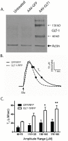
Figure 1. Characterization of AAV-GLT1 in vitro and in vivo.
(A) Western blot analysis for GLT-1 protein in HEK293 cells that were transfected with the AAV-GLT1 packaging plasmid for 24 hours. GLT-1 appears as two prominent bands 150 kD and 60 kD , . (B) Representative graphs showing time course of glutamate signals (amplitudes in 170–185 uM glutamate range) and the faster glutamate clearance rate in the striatum of animals injected with AAV-GLT1 compared to AAV-GFP. (C) Glutamate clearance (Tc) in the striatal hemisphere treated with AAV-GLT1 was significantly increased compared to the AAV-GFP -treated hemisphere. * p<0.05, ** p<0.01.
Figure 2
Figure 2. Transduction of rat cortex by three site injection of AAV-GFP serotype 1.
(A) Schematic representation of rat skull with injection sites (black dots). (B) Dashed line points to brain section cut in a sagittal plane showing 3 injections sites by GFP fluorescence (green;scale = 0.5 mm). (C) Coronal section of injection site showing expression of GFP from a dsAAV1eGFP injection. Coronal sections immunostained for neuronal marker, NeuN (red; D–F) or the astrocytic marker, GFAP (red;G–I) show that GFP (green) colocalizes to both neurons and astrocytes in the cortex. Arrowheads illustrate example of NeuN (D,E) or GFAP (G,H) double-labeled cells. Scale bar = 100 um.
Figure 3
Figure 3. AAV-GLT1 significantly reduces infarction size in region of viral injection.
Local administration of AAV-GLT1 reduced brain infarction. (A) AAV-GLT1 significantly reduced the maximal infarct area compared to AAV-GFP treated animals (p<0.01, Student's t-test). (B) Analysis of infarction based on 2 mm brain section showed a significant reduction in brain infarction in animals treated with AAV-GLT1 (p<0.01, Two-way ANOVA). Significant differences in the infarction area per slice were observed in slices 3 through 5 (arrows) which contain the viral injection sites (*p<0.05). (C) Representative 2 mm brain sections stained with TTC. Rats received 3 cortical injections of AAV-GLT1 or AAV-GFP into the right hemisphere.
Figure 4
Figure 4. AAV-GLT1 promotes behavioral recovery after cerebral ischemia.
Behavioral tests were carried out at 2, 8 and 14 days after the MCAo and analyzed with 2-way ANOVA. AAV-GLT1 injection significantly reduced body asymmetry in an elevated body swing test (A) and neurological abnormality scores in Bederson's test (B). *p<0.05, **p<0.01, ***p<0.001; Bonferroni test.
Figure 5
Figure 5. Pre-treatment with AAV-GLT1 reduces glutamate overflow in stroke rats.
Microdialysis was performed in the lesioned cortex. In animals receiving AAV-GFP, MCAo caused an increase in extracellular glutamate. In animals receiving AAV-GLT1, MCAo -induced glutamate overflow was significantly reduced (p<0.01, Two-way, AAV-GLT1 vs AAV-GFP post-MCAO;*p<0.05, **p<0.01, ***p<0.001; Bonferroni test.
Figure 6
Figure 6. Pre-treatment with AAV-GLT1 reduces TUNEL staining following MCAo.
TUNEL was examined at 24 hours after MCAo in the region of infarction near site of viral transduction. (A) Schematic of coronal secion near bregma indicating cortical region (box) of representative images in C–F. (B) AAV-GLT1 significantly reduced TUNEL staining in ischemic cortex compared to AAV-GFP (p<0.05, Student's t-test). Representative TUNEL from AAV-GFP (C) and AAV-GLT1(D). Cresyl violet staining in on adjacent brain sections from AAV-GFP (E) and AAV-GLT1(F) treated animals. Scale bar = 0.2 mm.

References

    1. Martin RL, Lloyd HGE, Cowan AI. The early events of oxygen and glucose deprivation - setting the scene for neuronal death. Trends in Neurosciences. 1994;17:251–257. - PubMed
    1. Doyle KP, Simon RP, Stenzel-Poore MP. Mechanisms of ischemic brain damage. Neuropharmacology. 2008;55:310–318. - PMC - PubMed
    1. Garber K. Stroke treatment–light at the end of the tunnel? Nat Biotechnol. 2007;25:838–840. - PubMed
    1. Sheardown MJ, Nielsen EO, Hansen AJ, Jacobsen P, Honore T. 2,3-Dihydroxy-6-Nitro-7-Sulfamoyl-Benzo(F)Quinoxaline - A neuroprotectant for cerebral ischemia. Science. 1990;247:571–574. - PubMed
    1. Simon RP, Swan JH, Griffiths T, Meldrum BS. Blockade of N-methyl-D-aspartate receptors may protect against ischemic damage in the brain. Science. 1984;226:850–852. - PubMed

Publication types

MeSH terms

Substances