. 2011;6(8):e22664.
doi: 10.1371/journal.pone.0022664. Epub 2011 Aug 11.

Characterization of the PRMT gene family in rice reveals conservation of arginine methylation

Affiliations

Characterization of the PRMT gene family in rice reveals conservation of arginine methylation

Ayaz Ahmad et al. PLoS One. 2011.

Abstract

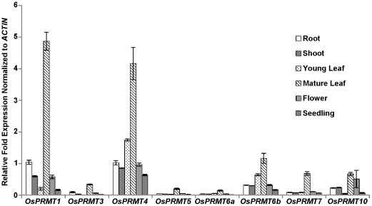
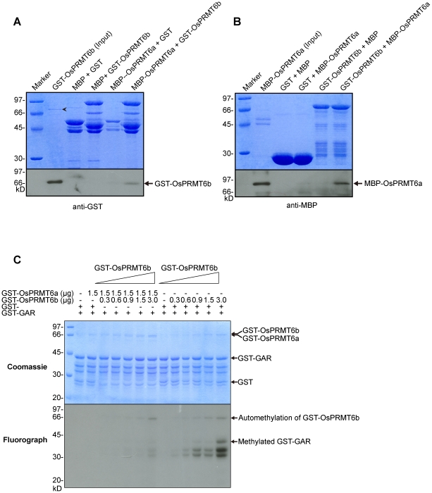
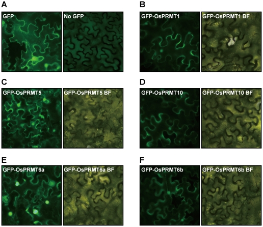
Post-translational methylation of arginine residues profoundly affects the structure and functions of protein and, hence, implicated in a myriad of essential cellular processes such as signal transduction, mRNA splicing and transcriptional regulation. Protein arginine methyltransferases (PRMTs), the enzymes catalyzing arginine methylation have been extensively studied in animals, yeast and, to some extent, in model plant Arabidopsis thaliana. Eight genes coding for the PRMTs were identified in Oryza sativa, previously. Here, we report that these genes show distinct expression patterns in various parts of the plant. In vivo targeting experiment demonstrated that GFP-tagged OsPRMT1, OsPRMT5 and OsPRMT10 were localized to both the cytoplasm and nucleus, whereas OsPRMT6a and OsPRMT6b were predominantly localized to the nucleus. OsPRMT1, OsPRMT4, OsPRMT5, OsPRMT6a, OsPRMT6b and OsPRMT10 exhibited in vitro arginine methyltransferase activity against myelin basic protein, glycine-arginine-rich domain of fibrillarin and calf thymus core histones. Furthermore, they depicted specificities for the arginine residues in histones H3 and H4 and were classified into type I and Type II PRMTs, based on the formation of type of dimethylarginine in the substrate proteins. The two homologs of OsPRMT6 showed direct interaction in vitro and further titrating different amounts of these proteins in the methyltransferase assay revealed that OsPRMT6a inhibits the methyltransferase activity of OsPRMT6b, probably, by the formation of heterodimer. The identification and characterization of PRMTs in rice suggests the conservation of arginine methylation in monocots and hold promise for gaining further insight into regulation of plant development.


Conflict of interest statement

Competing Interests: The authors have declared that no competing interests exist.

Figures

Figure 1
Figure 1. Members of the protein arginine methyltransferase family of proteins in Oryza sativa.
Eight members of the Oryza sativa PRMT family; sharing the conserved PRMT signature motifs I, post-I, -II, -III, THW loop (Black bars) and double E loop (grey bars). PRMT3 harbors an additional Zn Finger domain (lined bar).
Figure 2
Figure 2. Relative expression patterns of the OsPRMTs.
Total RNAs were isolated from plant tissues; root, young leaf, mature leaf, shoot, flower and whole seedling of the Nipponbare strain and cDNAs were synthesized. The relative fold change in the expression of the OsPRMT1, OsPRMT3, OsPRMT4, OsPRMT5, OsPRMT6a, OsPRMT6b, OsPRMT7 and OsPRMT10 was normalized to the expression of ACTIN. The error bars represent standard deviation.
Figure 3
Figure 3. In vitro methyltransferase activity assay of the OsPRMTs.
Methyltransferase activity assay of OsPRMT1 (A), OsPRMT4 (B), OsPRMT5 (C), OsPRMT6a (D), OsPRMT6b (E), and OsPRMT10 (F). GST and GST-OsPRMTs were bound to the GST beads and incubated with indicated substrates; myelin basic protein, GST-GAR and calf thymus core histones in the presence of [3H]SAM for 3 hours in a final volume of 30 µl of HMT buffer (20 mM Tris/HCl, 4 mM sodium EDTA, 1 mM PMSF and 1 mM dithiothreitol). Methylated proteins were separated by 10% SDS-PAGE, stained with Coomassie blue (upper panels), destained, soaked in amplify (Amersham Biosciences, NAMP100) dried and visualized by fluorography by exposing to the x-ray film at −80°C for 48 hours (lower panels). The corresponding proteins are indicated above the upper panels. The arrow “→” points GST-Tagged full length OsPRMTs whereas the asteric “*” represents the substrate proetins i.e. GST-GAR, myelin basic protein and core histones (H3, H4, H2A and H2B). The arrow head “formula image
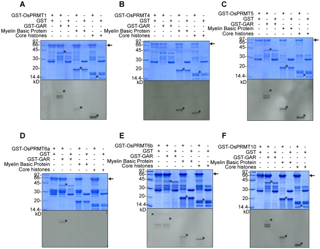
” indicates the automethylation of OsPRMT6b.
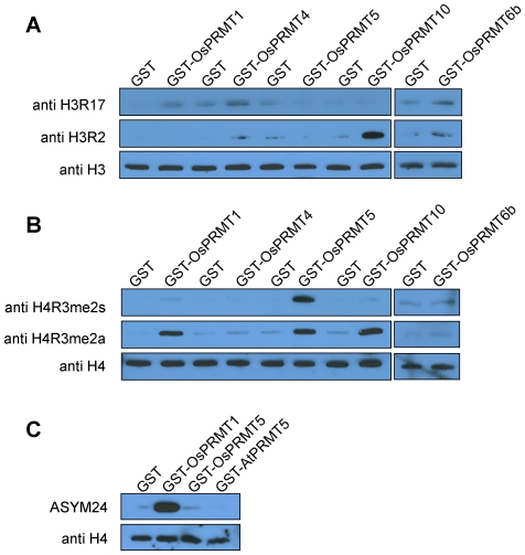
Figure 4
Figure 4. Site specificities of the OsPRMT1, OsPRMT5, OsPRMT6b, and OsPRMT10.
(A, B and C) Purified calf thymus core histones were either incubated with negative control, GST or GST-OsPRMT1, GST-OsPRMT5, GST-OsPRMT6b, GST-OsPRMT10 and GST-AtPRMT5 and were separated by 15% SDS-PAGE, followed by western blot analysis using anti-H3R2me2a, anti-H3R17me2a, anti-H4R3me2a, anti-H4R3me2s, and ASYM24 antibodies. An equal amount of the reaction mixtures were separated by the 15% SDS-PAGE, followed by western blot analysis using anti-H3 and anti-H4 antibodies, for equal loading (lowest panels in A, B and C). The antibodies are indicated to the left of each panel, whereas the corresponding PRMTs are represented above the upper panels.
Figure 5
Figure 5. OsPRMT6a and OsPRMT6b directly interact in vitro.
(A and B) GST, MBP, MBP-OsPRMT6a and GST-OsPRMT6b were expressed in E. coli. Reciprocal pull-down assay was performed as described in the experimental part. Equal amount of each mixture of proteins was separated on two separate 10% SDS-PAGE, one for Coomassie staining (upper panels A and B) for equal loading and the other for western blotting (lower panels A and B). GST and MBP alone were used as negative controls. The protein pairs are indicated at the top of the upper panels. MBP-OsPRMT6a served as bait and GST-OsPRMT6b as prey (A) and vice versa (B). A 10 µl cell extract from E. coli containing MBP-OsPRMT6a and GST-OsPRMT6b was used as input. Western blotting using GST and MBP monoclonal antisera revealed the interaction between OsPRMT6a and OsPRMT6b. The arrow head “???” indicates MBP-OsPRMT6a. (C) 0.3, 0.6, 0.9, 1.5 and 3.0 µg of GST-OsPRMT6b was mixed either with 1.5 µg of GST-OsPRMT6a or with GST protein alone (as control) in an in vitro methyltransferase activity assay as described in figure 3. The Coomassie blue stained and dried gel was exposed at −80°C for four days.
Figure 6
Figure 6. Subcellular localization of the OsPRMT1, OsPRMT5, OsPRMT6a, OsPRMT6b, and OsPRMT10.
OsPRMT1, OsPRMT5, OsPRMT6a, OsPRMT6b, and OsPRMT10 were fused in frame to GFP in binary vector, transformed to the Agrobactrial strain, EHA105 and injected into the Nicotiana benthamiana leaves. Expression of GFP and GFP-tagged OsPRMT1, OsPRMT5, OsPRMT6a, OsPRMT6b, and OsPRMT10 was observed under fluorescence microscopy and bright field microscopy (designated by BF) after 2–3 days.

References

    1. Wolf SS. The protein arginine methyltransferase family: an update about function, new perspectives and the physiological role in humans. Cell Mol Life Sci. 2009;66:2109–2121. - PMC - PubMed
    1. Pahlich S, Zakaryan RP, Gehring H. Protein arginine methylation: Cellular functions and methods of analysis. Biochem Biophys Acta. 2006;1764:1890–1903. - PubMed
    1. Krause CD, Yang ZH, Kim YS, Lee JH, Cook JR, et al. Protein arginine methyltransferases: evolution and assessment of their pharmacological and therapeutic potential. Pharmacol Ther. 2007;113:50–87. - PubMed
    1. Bedford MT. Arginine methylation at a glance. J Cell Sci. 2007;120:4243–4246. - PubMed
    1. Gary JD, Clarke S. RNA and protein interactions modulated by protein arginine methylation. Prog Nucleic Acid Res Mol Biol. 1998;61:65–131. - PubMed

Publication types

MeSH terms