. 2011;6(8):e22880.
doi: 10.1371/journal.pone.0022880. Epub 2011 Aug 11.

Single molecule analysis of c-myb alternative splicing reveals novel classifiers for precursor B-ALL

Affiliations

Single molecule analysis of c-myb alternative splicing reveals novel classifiers for precursor B-ALL

Ye E Zhou et al. PLoS One. 2011.

Abstract

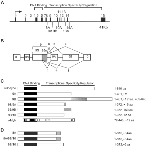
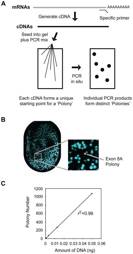
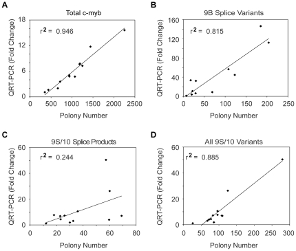
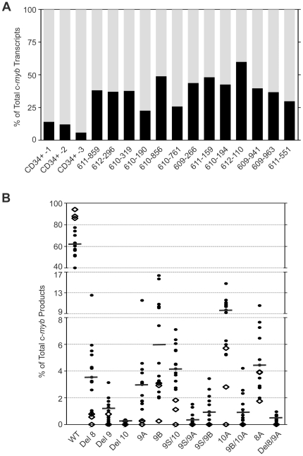
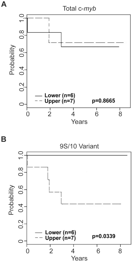
The c-Myb transcription factor, a key regulator of proliferation and differentiation in hematopoietic and other cell types, has an N-terminal DNA binding domain and a large C-terminal domain responsible for transcriptional activation, negative regulation and determining target gene specificity. Overexpression and rearrangement of the c-myb gene (MYB) has been reported in some patients with leukemias and other types of cancers, implicating activated alleles of c-myb in the development of human tumors. Alternative RNA splicing can produce variants of c-myb with qualitatively distinct transcriptional activities that may be involved in transformation and leukemogenesis. Here, by performing a detailed, single molecule assay we found that c-myb alternative RNA splicing was elevated and much more complex in leukemia samples than in cell lines or CD34+ hematopoietic progenitor cells from normal donors. The results revealed that leukemia samples express more than 60 different c-myb splice variants, most of which have multiple alternative splicing events and were not detectable by conventional microarray or PCR approaches. For example, the single molecule assay detected 21 and 22 splice variants containing the 9B and 9S exons, respectively, most of which encoded unexpected variant forms of c-Myb protein. Furthermore, the detailed analysis identified some splice variants whose expression correlated with poor survival in a small cohort of precursor B-ALL samples. Our findings indicate that single molecule assays can reveal complexities in c-myb alternative splicing that have potential as novel biomarkers and could help explain the role of c-Myb variants in the development of human leukemia.


Conflict of interest statement

Competing Interests: The authors have declared that no competing interests exist.

Figures

Figure 1
Figure 1. Alternative splicing in the c-myb gene.
(A) Structure of the human c-myb gene. The diagram shows the exon structure of the human c-myb gene, which spans more than 41,000 nucleotides. The arrow marks the major transcription initiation site and exons 1–15, which are included in the wild type version of c-Myb, are indicated above the line. The highly conserved DNA binding domain of c-Myb is encoded by exons 4–6, and the C-terminal domains that control transcriptional activity and specificity are encoded by exons 7–15, as indicated. The latter region also includes the 6 alternative exons, shown below the line. (B) Alternative splicing in exons 8–10. A cryptic splice donor site in exon 9 generates a short (9S) version lacking 85 nt (shaded). Both the long and short forms of exon 9 can be spliced to alternative exons 9A or 9B or to exon 10. (C) Diagrams of wild type c-Myb and the 9A, 9B, 9S/9A, 9S/9B and 9S/10 splice variants. The structure of the v-Myb protein encoded by Avian Myeloblastosis Virus is shown for comparison. The highly conserved Myb DNA binding domain is shaded black. The variant proteins lack the normal C-terminus of c-Myb and have unique regions, indicated by shaded boxes. The numbers on the right summarize the structures of the proteins with the included amino acids (aa) and the changes relative to wild type c-Myb. (D) Structures of the proteins encoded by splice variants 8A, 8A/9S/10 and 9S/10. These variants are described in the text. Labels and numbering are as in panel (C).
Figure 2
Figure 2. Sensitivity and linearity of the polony assay.
(A) Outline of polony assay. Gene-specific primers are used to convert mRNA to cDNA, which is then seeded into a thin acrylamide gel containing primers, enzymes and other components necessary for in situ PCR, which results in the formation of “PCR colonies”, or polonies. The polonies are fixed in the gel, but can be interrogated by sequential hybridization with specific fluorescently labeled oligonucleotides. (B) Sensitivity of polony assay. Plasmids containing cDNAs for wild type c-myb or the 8A splice variant were mixed in a ratio of 5,000∶1. After in situ PCR, the slide was hybridized sequentially with fluorescently labeled probes specific for exon 8, exon 8A or exon 11. The full polony slide is shown at left and the enlargement at right shows a single detected exon 8A-containing splice variant (white). In the false color image the polonies containing only exons 8 and 11 appear Cyan in color, the polony containing all three exons appears white. (C) Linearity of polony assay. Different amounts of wild-type c-myb plasmid were used as template in the polony assay and the number of polonies detected with the exon 11 probe was plotted against amount of plasmid template used in the assay. The assay was very linear, as shown by the correlation coefficient r2, which was 0.99.
Figure 3
Figure 3. Comparison of exon-specific (QPCR) to single molecule (polony) assays.
Scatter plots compare the levels of different c-myb splice variants detected by bulk QPCR or single molecule polony-based assays for (A) total c-myb transcripts or (B–C) the 9B or 9S/10 splice variants, respectively. The QPCR and polony assays agree nicely (r2 = 0.946) for total c-myb transcripts, but the results for the 9B variant agree only modestly (r2<0.82) and the results for the 9S/10 variant did not correlate well (r2<0.25). (D) Compiling the polony data by combining all the detected transcripts that contain the 9S/10 exon produces data that correlate much better (r2>0.88) with the QPCR data. Thus the differences in the two assays are due to the exclusion of variants in the polony results that are predicted to produce different protein products, which are detectable in the polony assay but not by QPCR.
Figure 4
Figure 4. Alternative splicing of c-myb transcripts in normal and leukemic cells.
(A) Rates of c-myb alternative splicing in normal cells and leukemias. Polony assays were used to measure the levels of normal and alternatively spliced c-myb transcripts in normal CD34+ progenitors from healthy donors (n = 3) or in pediatric precursor B-ALL patient samples (n = 13). The black and gray sections of each bar indicate the fractions of alternatively spliced or wild type transcripts, respectively. (B) Patterns of c-myb alternative splicing in pediatric precursor B-ALL samples. Dots indicate levels of wt or splice variant expression, as a fraction of total c-myb transcripts detected, as determined by polony assay for pediatric precursor B-ALL samples (n = 13, filled circles) or CD34+ cells from normal donors (n = 3, empty diamonds). Each symbol represents the average of duplicate measurements and the horizontal bars represent the median values for the leukemia samples.
Figure 5
Figure 5. Survival plots for patients grouped by c-myb variant expression levels.
Precursor B-ALL patients (n = 13) were grouped according to their expression above (dotted line) or below (solid line) the median expression level for (A) total c-myb transcripts or (B) the 9S/10 variant. The plots show overall survival curves for the high expression or low expression groups of leukemia samples, with p-values as indicated.

References

    1. Mucenski ML, McLain K, Kier AB, Swerdlow SH, Schreiner CM, et al. A functional c-myb gene is required for normal murine fetal hepatic hematopoiesis. Cell. 1991;65:677–689. - PubMed
    1. Sandberg ML, Sutton SE, Pletcher MT, Wiltshire T, Tarantino LM, et al. c-Myb and p300 regulate hematopoietic stem cell proliferation and differentiation. Dev Cell. 2005;8:153–166. - PubMed
    1. Sakamoto H, Dai G, Tsujino K, Hashimoto K, Huang X, et al. Proper levels of c-Myb are discretely defined at distinct steps of hematopoietic cell development. Blood. 2006;108:896–903. - PubMed
    1. Fahl SP, Crittenden RB, Allman D, Bender TP. c-Myb is required for pro-B cell differentiation. J Immunol. 2009;183:5582–5592. - PMC - PubMed
    1. Hu T, Simmons A, Yuan J, Bender TP, Alberola-Ila J. The transcription factor c-Myb primes CD4+CD8+ immature thymocytes for selection into the iNKT lineage. Nat Immunol. 2010;11:435–441. - PMC - PubMed

Publication types

MeSH terms

LinkOut - more resources