. 2011;6(8):e22960.
doi: 10.1371/journal.pone.0022960. Epub 2011 Aug 11.

A role for glutamate transporters in the regulation of insulin secretion

Affiliations

A role for glutamate transporters in the regulation of insulin secretion

Runhild Gammelsaeter et al. PLoS One. 2011.

Abstract

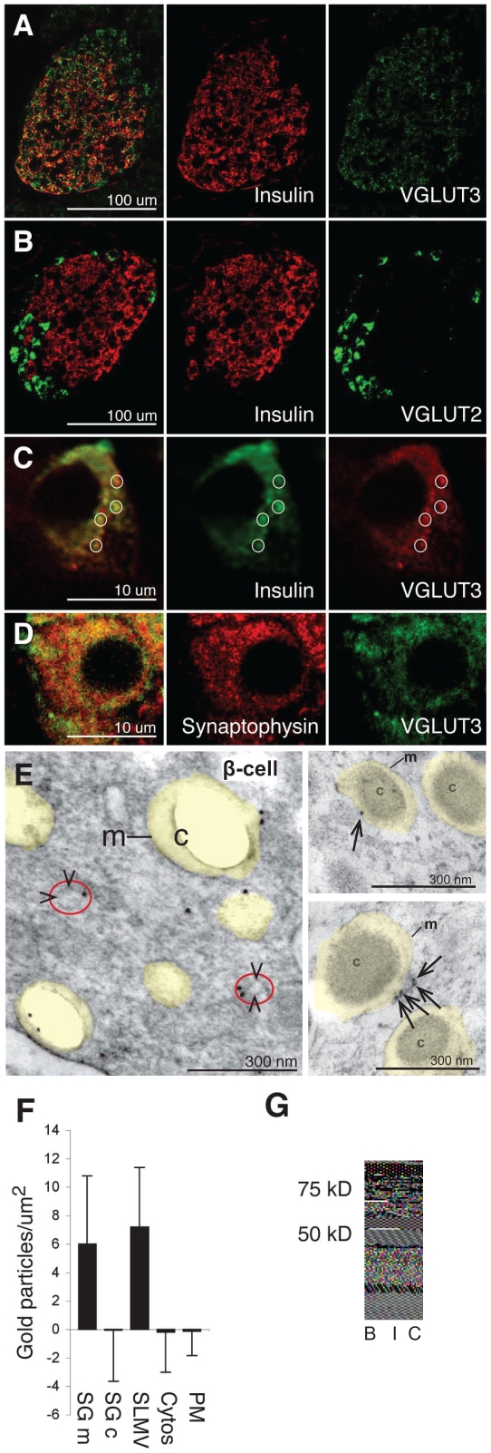
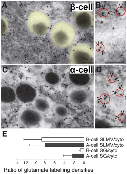
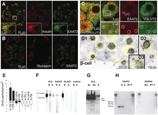
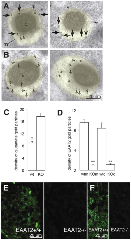
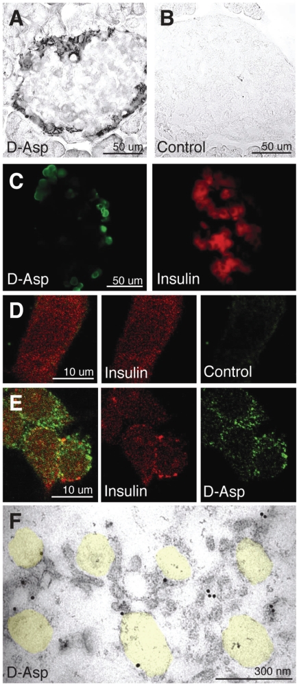
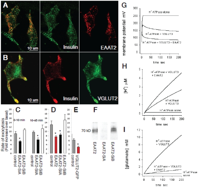
In the brain, glutamate is an extracellular transmitter that mediates cell-to-cell communication. Prior to synaptic release it is pumped into vesicles by vesicular glutamate transporters (VGLUTs). To inactivate glutamate receptor responses after release, glutamate is taken up into glial cells or neurons by excitatory amino acid transporters (EAATs). In the pancreatic islets of Langerhans, glutamate is proposed to act as an intracellular messenger, regulating insulin secretion from β-cells, but the mechanisms involved are unknown. By immunogold cytochemistry we show that insulin containing secretory granules express VGLUT3. Despite the fact that they have a VGLUT, the levels of glutamate in these granules are low, indicating the presence of a protein that can transport glutamate out of the granules. Surprisingly, in β-cells the glutamate transporter EAAT2 is located, not in the plasma membrane as it is in brain cells, but exclusively in insulin-containing secretory granules, together with VGLUT3. In EAAT2 knock out mice, the content of glutamate in secretory granules is higher than in wild type mice. These data imply a glutamate cycle in which glutamate is carried into the granules by VGLUT3 and carried out by EAAT2. Perturbing this cycle by knocking down EAAT2 expression with a small interfering RNA, or by over-expressing EAAT2 or a VGLUT in insulin granules, significantly reduced the rate of granule exocytosis. Simulations of granule energetics suggest that VGLUT3 and EAAT2 may regulate the pH and membrane potential of the granules and thereby regulate insulin secretion. These data suggest that insulin secretion from β-cells is modulated by the flux of glutamate through the secretory granules.


Conflict of interest statement

Competing Interests: The authors have declared that no competing interests exist.

Figures

Figure 1
Figure 1. The vesicular glutamate transporter VGLUT3 is localized in secretory granules and synaptic-like microvesicles (SLMVs) in pancreatic β-cells.
(A) VGLUT3 (green) co-localizes with insulin (red), but is also found in peripheral non-B islet cells. (B) VGLUT2 (green) does not co-localize with insulin (red). (C) A single β-cell from the islet presented in panel A: VGLUT3 (red) co-localizes partly with insulin (green). The circles highlight some of the overlapping VGLUT3 and insulin dots. (D) A single β-cell: VGLUT3 (green) co-localizes partly with synaptophysin (red) in small dots. (E) Electron micrograph showing immunogold particles for VGLUT3 in two β-cells. Secretory granules are indicated by transparent yellow. m, membranes of secretory granules. c, core of the secretory granule (of which some disappeared in the preparation procedure). SLMVs are indicated by arrowheads and red circles. (F) Quantification of VGLUT3 in β-cells (n = 8 cells). Immunogold particle densities (mean number of gold particles/µm2 ±SD) in the membrane of the granules (SGm) and in SLMVs are significantly higher than in the core of the β-cell granules (SGc), cytosol and plasma membranes (PM) (background labeling, quantified over mitochondria, is subtracted, see Methods) (p<0.001, Mann-Whitney-U test, two tails). (G) Western blots of rat brain tissue (B) and isolated rat islets (I) probing for VGLUT3. (C), islet blot without the VGLUT3 primary antibodies.
Figure 2
Figure 2. The glutamate concentration in SLMVs is much higher than in secretory granules, and lower in β-cell secretory granules than in α-cell secretory granules.
(A) Immunogold particles representing glutamate in β-cell cytoplasm are scarce over secretory granules (transparent yellow). (B) Close up showing glutamate gold particles in β-cell SLMVs (arrowheads, red circle). (C) Immunogold particles representing glutamate in α-cell cytoplasm. (D) Close up showing glutamate gold particles in α-cell SLMVs (arrowheads, red circle). Scale bars A–D, 100 nm. (E) Immunogold quantification shows that the secretory granule (SG)/cytosol (cyto) ratio (mean±SD) of net glutamate labelling (background subtracted, see Methods) is significantly lower in β-cells than in α-cells (p<0.05, n = 5 cells of each kind) and that the SLMV/cytosol ratio is much higher than the SG/cytosol ratio in both α- and β-cells (p<0.01, n = 5 cells of each kind) (Mann-Whitney-U test, two tails). The mean glutamate labelling density (average number of gold particles/µm2±SD) in α-cell granules was 33.6±14.9, whereas the value in α-cell cytosol was 21.1±4.6 (p<0.05, Mann-Whitney-U test, two tails). In β-cells the glutamate densities were 19.7±12.4 in the granules and 30.2±7.6 in the cytosol (p<0.05, Mann-Whitney-U test, two tails). From ultrathin test sections with conjugates containing known concentrations of glutamate, which were processed with the glutamate antibodies in parallel with the glutamate labelling of islet tissue, a relationship between the concentration of fixed glutamate and the gold particle density in islet tissue can be approximately estimated. In β-cells the approximate concentration of glutamate was estimated to be in the lower mM range (2–3 mM in secretory granules and cytosolic matrix, respectively).
Figure 3
Figure 3. The glutamate transporter EAAT2 is selectively localized in β-cell secretory granules.
(A–B) EAAT2 (green) co-localizes with insulin (red in A) in β-cells but not with glucagon in α-cells (red B). (C) EAAT2 (red) co-localizes partly with the vesicular glutamate transporter VGLUT3 (green). At higher magnification (right panels) it is evident that there are granules containing both EAAT2 and VGLUT3, some of which are indicated by circles. (D1–D3) Electron micrographs showing that EAAT2 immunogold particles are localized in secretory granules (transparent yellow) in three different β-cells. m, granule membrane. c, granule core. Arrowheads (/\), plasma membrane. E, Quantification of EAAT2 in different cellular compartments in α- and β-cells. The values are mean number of EAAT2 gold particles/µm2±SD in the various tissue compartments in 7 α- and 7 β-cells. The EAAT2 density is significantly higher in the membrane (B SG m) and core (B SG c) of the granules in β-cells, than in the cytosol of α- and β-cells (A cyto and B cyto), the plasma membrane of α- and β-cells (A PM and B PM) and the core (A SG c) and limiting membrane (A SG m) of α-cell granules (p<0.001, Mann-Whitney-U test, two tails). (Background labelling was subtracted, see Methods. Except for B SG m and B SG c, all other compartments observed were at background levels.) The quantitative data presented are from one animal and similar results were obtained in two other animals. F, Western blots of isolated rat pancreatic islets (I) and rat brain tissue (B) using EAAT2 antibodies raised against both the C-terminal (B12) and the N-terminal (monoclonal (M)) parts. EAAT1 showed no band in islet tissue. Controls (C) without primary antibody showed no band. Note that the Westerns were run with several different homogenate concentrations, so that the lanes to be directly compared could mostly not be on the same membrane, however all membranes were run in the same set of experiments. (G) Western blot of isolated rat pancreatic islets (I), and rat brain tissue (2 separate brains, B1 and B2) all on the same membrane using EAAT2 antibodies raised against the EAAT2 C-terminal (B12). (H) RT-PCR of rat brain tissue (B) and isolated islets (I) in which there was a PCR signal for EAAT2 at about 100 bp (expected value), but not for human GAPDH.
Figure 4
Figure 4. Lack of EAAT2 and increased concentration of glutamate in secretory granules in EAAT2 KO mice.
(A–B) Immunogold electron micrographs showing EAAT2 (large gold particles, long arrows) and glutamate (small gold particles, short arrows) in β-cell secretory granules (transparent yellow) in wt (A) and KO (B) islets (two examples of secretory granules shown for each phenotype). m, granule membrane. c, granule core. (C–D) Quantitative representation of the glutamate (C) and EAAT2 (D) gold particle densities in β-cell secretory granules in wild type (wt) and knock out (KO) mice. The values are mean numbers of gold particles/µm2±SEM in 4 KO and 4 wt animals (background subtracted, see Methods). 119 secretory granules in wt mice and 179 secretory granules in KO mice were included in the quantifications. *, the value in KO secretory granules is significantly higher than the value in wt secretory granules (p<0.01, Mann-Whitney-U test, two tails) and **, the values in the limiting membrane (m) and the core (c) of the secretory granules are significantly lower in KO than in wt mice (p<0.01, Mann-Whitney-U test, two tails). (E–F) Islet (E) and brain (F) tissue from wild type (EAAT2+/+) and EAAT2 KO (EAAT2−/−) mice labelled with the EAAT2 antibodies.
Figure 5
Figure 5. The glutamate analogue D-aspartate is neither taken up through the plasma membrane in intact β-cells nor into SGs in permeabilized β-cells.
A–C, Acutely prepared slices of islet tissue were incubated with exogenous D-aspartate (100 µM) before aldehyde fixation and labelling with antibodies that selectively recognize D-aspartate. (A–B) Immunoperoxidase labelling shows that the tissue not exposed to D-aspartate (Control) is unlabelled, while in islets exposed to D-aspartate (D-Asp) labelling is observed only in the peripheral α-cell area, not in the central β-cell area of the islet. (C) Immunofluorescence shows that the central insulin positive β-cells are negative for exogenous D-aspartate and that the peripheral non-insulin α-cells are labelled. (D–F) Streptolysin-O permeabilized INS-1E cells were exposed to different concentrations of exogenous D-aspartate (0–3 mM) before fixation and labelling with the D-aspartate antibodies. (D) In cells not exposed to D-aspartate (Control) there was no labelling for D-aspartate, only for insulin (red). (E) In cells exposed to 1 mM D-aspartate staining with the D-aspartate antibodies (green) is observed. There was some weak co-localization (yellow) with insulin (red) that is attributable to extra granular fixation of D-aspartate (see F). (F) Electron micrograph of permeabilized INS cells exposed to 1 mM D-aspartate shows no significant D-aspartate labelling inside the secretory garnules (indicated in transparent yellow). Note some labelling along the limiting membrane of secretory granules and in the cytosol, reflecting fixation of exogenous D-aspartate to extragranular proteins.
Figure 6
Figure 6. The effect of EAAT2 and VGLUT expression on secretory granule energetics and exocytosis.
(A) INS-1E cells were transfected with EAAT2-c-Myc and the EAAT2 protein was visualized with anti-c-Myc antibodies (red). EAAT2 co-localized with insulin (green). (B) INS-1E cells were transfected with GFP-VGLUT2 and VGLUT2 visualized by the fluorescence of the GFP tag. VGLUT2 (green) co-localized with insulin (red). (C) INS-1E cells were transiently co-transfected with a plasmid encoding hGH and with vectors directing the sequence of two different short interfering RNAs for EAAT2 (EAAT2 SiA and EAAT2 SiB) or with empty pSUPER vector (control). The cells were incubated under basal and stimulatory conditions (see methods) for 10 and 45 min. hGH secretion was measured by ELISA as the fold increase over basal conditions. The EAAT2 SiA values were significantly different from control and EAAT2 SiB values (*, unpaired Student's t-test, p<0.05, n = 3). (D) In another set of experiments INS-1E cells were cotransfected with hGH and the pSUPER control vector alone (control) or with EAAT2, EAAT2 SiA or EAAT2 SiB, and incubated for 45 min as in C. EAAT2 and EAAT2 SiA values were significantly different from control and EAAT2 SiB values (*, unpaired Student's t-test, p<0.05, n = 3). (E) INS-1E cells were transiently co-transfected with hGH and VGLUT2-GFP or GFP (control). The VGLUT2 value was significantly different from the control value (*, unpaired Student's t-test, p<0.05, n = 3). (F) INS-1E cells were cotransfected with c-Myc-tagged EAAT2 and the two siRNAs targeting different sequences of EAAT2 (EAAT2 SiA and EAAT2 SiB). The Western blots show that the EAAT2 SiA almost completely blocked the expression of EAAT2, whereas EAAT2 SiB had no effect. (G–I) Simulations of the effect of VGLUT3 and EAAT2 on granule energetics. Granule cell membrane potential (G), [H+] (H) and [glutamate] (I) are shown for a granule membrane containing the H+-ATPase alone, ATPase and VGLUT3, or ATPase, VGLUT3 and EAAT2 (see supplementary text S1).

References

    1. Ottersen OP, Storm-Mathisen J. Glutamate- and GABA-containing neurons in the mouse and rat brain, as demonstrated with a new immunocytochemical technique. J Comp Neurol. 1984;229:374–392. - PubMed
    1. Fremeau RT, Jr, Voglmaier S, Seal RP, Edwards RH. VGLUTs define subsets of excitatory neurons and suggest novel roles for glutamate. Trends Neurosci. 2004;27:98–103. - PubMed
    1. Fremeau RT, Jr, Burman J, Qureshi T, Tran CH, Proctor J, et al. The identification of vesicular glutamate transporter 3 suggests novel modes of signaling by glutamate. Proc Natl Acad Sci U S A. 2002;99:14488–93. - PMC - PubMed
    1. Danbolt NC. Glutamate uptake. Prog Neurobiol. 2001;65:1–105. - PubMed
    1. Balcar V, Johnston GA. The structural specificity of the high affinity uptake of L-glutamate and L-aspartate by rat brain slices. J Neurochem. 1972;19:2657–66. - PubMed

Publication types

MeSH terms

Substances