. 2011;6(8):e23108.
doi: 10.1371/journal.pone.0023108. Epub 2011 Aug 11.

Pronounced hypoxia in models of murine and human leukemia: high efficacy of hypoxia-activated prodrug PR-104

Affiliations

Pronounced hypoxia in models of murine and human leukemia: high efficacy of hypoxia-activated prodrug PR-104

Juliana Benito et al. PLoS One. 2011.

Abstract

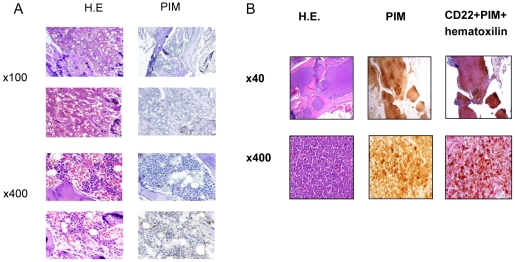
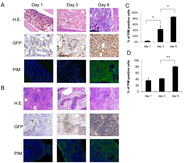
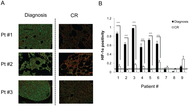
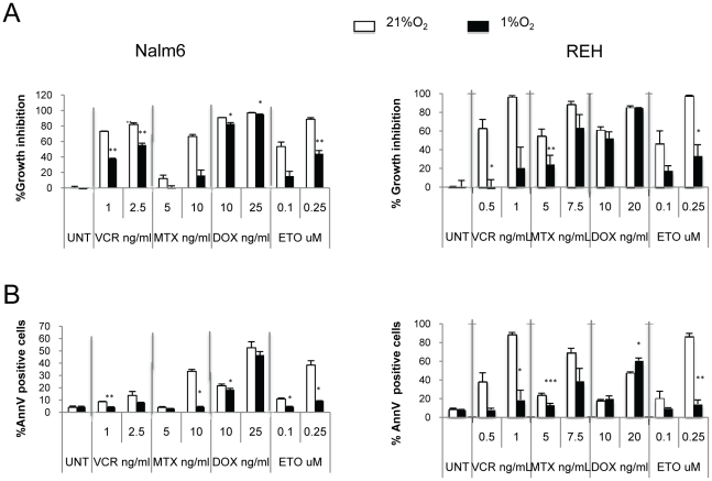
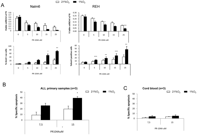
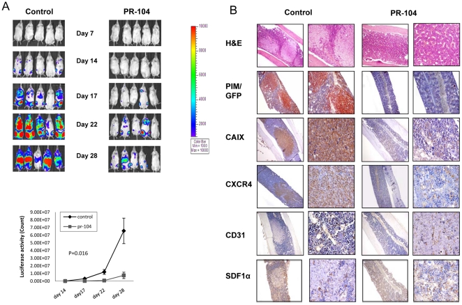
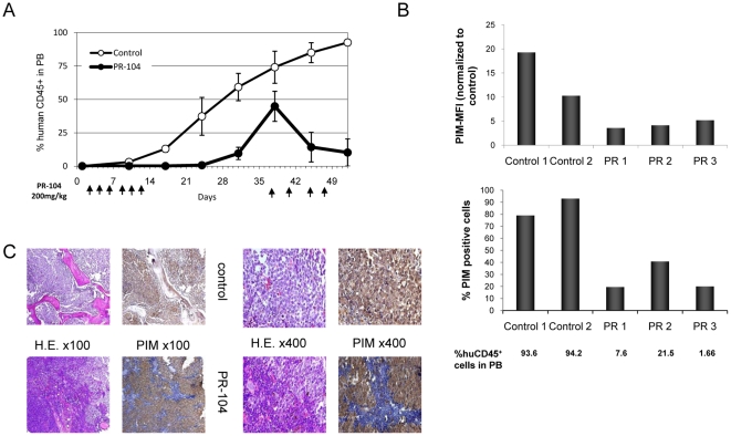
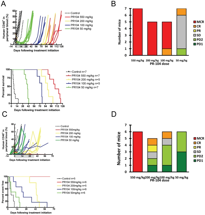
Recent studies indicate that interactions between leukemia cells and the bone marrow (BM) microenvironment promote leukemia cell survival and confer resistance to anti-leukemic drugs. There is evidence that BM microenvironment contains hypoxic areas that confer survival advantage to hematopoietic cells. In the present study we investigated whether hypoxia in leukemic BM contributes to the protective role of the BM microenvironment. We observed a marked expansion of hypoxic BM areas in immunodeficient mice engrafted with acute lymphoblastic leukemia (ALL) cells. Consistent with this finding, we found that hypoxia promotes chemoresistance in various ALL derived cell lines. These findings suggest to employ hypoxia-activated prodrugs to eliminate leukemia cells within hypoxic niches. Using several xenograft models, we demonstrated that administration of the hypoxia-activated dinitrobenzamide mustard, PR-104 prolonged survival and decreased leukemia burden of immune-deficient mice injected with primary acute lymphoblastic leukemia cells. Together, these findings strongly suggest that targeting hypoxia in leukemic BM is feasible and may significantly improve leukemia therapy.


Conflict of interest statement

Competing Interests: The authors have read the journal's policy and have the following conflicts: Dr. William R. Wilson holds stock in PROACTA Inc. and is a consultant to the company. This does not alter the authors' adherence to all the PLoS ONE policies on sharing data and materials.

Figures

Figure 1
Figure 1. Expansion of the hypoxic BM niche in an ALL xenograft model.
Representative images from one healthy control mouse (two different BM fields, A) or mice transplanted with CD22-expressing leukemic cells from an infant with MLL-rearranged B-lineage ALL (B). After 52 days, mice were injected with pimonidazole (PIM) 3 hrs prior to sacrifice. Areas of hypoxia were detected by PIM antibody, and leukemic cells by anti-CD22. Original magnification is shown next to each panel.
Figure 2
Figure 2. Expansion of the hypoxic BM niche in a syngenic model of blastic phase CML.
1×10⁷ GFP/YFP labeled cells expressing the oncogenes BCR/ABL and Nup98 were FACS-sorted and transplanted into irradiated (4.5GY) C57B16/J mice. At the indicated time-points, mice were injected with pimonidazole 3 hrs prior to sacrifice. Areas of hypoxia were detected by PIM, and leukemic cells by anti-GFP in BM (A) or spleen (B). (C) and (D) Quantification of PIM positive cells in BM and spleen, respectively by CRi spectral imaging and Inform software analysis (at least 3 mice per group were analyzed). Original magnification, ×100. *P<0.05; **P<0.01.
Figure 3
Figure 3. Expansion of the hypoxic BM niche in primary leukemia samples reverted following complete remission (CR).
HIF-1α was detected by IHC in BM biopsies from 9 ALL patients at diagnosis and at CR after induction chemotherapy. (A) Representative images from 3 patients are shown. Original magnification, ×500. (B) Quantification of HIF-1α expression using Cri Inform software. A threshold was established based on the scores of three normal BM samples (continuous line represents average HIF-1α and dashed line correspond to SD for these 3 samples, respectively) CR; complete remission *** P<0.0001, error bars SEM.
Figure 4
Figure 4. Hypoxia promotes resistance of leukemia cell lines to chemotherapy.
Cytotoxic effect of chemotherapy under normoxic and hypoxic conditions. Nalm6, REH ALL cells were treated with various chemotherapy drugs for 48 hours at 21% or 1% O2. Percentage of growth inhibition was calculated based on cell number. A and B: cell number and percentage of Annexin V–positive cells were determined by FACS. Error bars, SEM. * P<0.05; ** P<0.01; *** P<0.001.
Figure 5
Figure 5. In vitro hypoxia cytotoxicity of PR-104A.
(A) B-cell ALL cell lines Nalm6 and REH B-ALL were treated with PR-104A under normoxic or hypoxic conditions for 6 hours. Cell death, measured as percentage of Annexin V (AnnV)–positive cells and cell number were measured 48 hours later by FACS. (B) and (C) Three primary ALL samples (B) or three control CB (C) were treated with PR-104A at normoxic or hypoxic conditions for 6 hours. Cell death, measured as percentage of Specific Apoptosis [% Specific Apoptosis = (%AnnV sample-%AnnV control)/(100-%AnnV control)*100] were measured 24 hours later by FACS. *P<0.05; **P<0.01; ***P<0.0001.
Figure 6
Figure 6. PR-104 administration resulted in decreased tumor burden and hypoxic areas in a Nalm6/GFP ALL model.
NSG mice injected with Nalm6-GFP/Luciferase cells were treated with PR-104 starting on day 3 after injection (250 mg/kg, i.p. 3 times a week for 2 weeks). PIM was administered 3 hours prior to sacrifice. (A) Luciferase activity in PR-104-treated and control mice over the course of the experiment. Bottom graph: Luciferase quantification of control and PR-104 treated mice. (B) BM sections from control and treated mice stained for the hypoxia markers PIM and CAIX and for GFP, CXCR4, CD31 and SDF1α. Original magnification, ×100 and ×400 (H&E) ×40 and ×400 (PIM/GFP, CAIX, CXCR4, CD31 and SDF1α).
Figure 7
Figure 7. PR-104 induced decrease of circulating CD45+ cells and of hypoxia in NSG mice injected with a primary B-lineage ALL sample.
(A) Time course of changes in percentage of circulating CD45+ cells determined in peripheral blood by FACS (n = 9/group). Arrows at the bottom indicate days when PR-104 was administered. (B) PIM in BM flushes from two control and three PR-104-treated mice determined by FACS. PIM was administered 3 hour prior to sacrifice on Day 52. PIM MFI is plotted as a ratio between the sample MFI/unstained control MFI. Bottom panel: percentage of PIM positive cells. The % of human CD45 positive cells in peripheral blood (PB) is indicated for each mouse. (C) Pimonidazole adducts and H&E staining in BM sections from control and PR-104-treated mice determined by IHC. Original magnification is indicated.
Figure 8
Figure 8. Dose-response efficacy of PR-104 against ALL-8 and ALL-19 in vivo.
(A and C) The responses of NOD/SCID mice engrafted with ALL-8 (A) or ALL-19 (C) xenograft to treatment with PR-104 at decreasing doses are represented as percentages of human CD45+ cells in the peripheral blood of individual mice over time. PR-104 was administered once a week for 6 weeks via i.p. injection; the shaded area corresponds to the duration of treatment. Kaplan-Meier plots of EFS, from which LGD was calculated (difference between median EFS of treated and control mice) and compared using the logrank test. The corresponding values are shown in Table S1. (B and D) Distribution of objective response measures for ALL-8 (B) or ALL-19 (D) engrafted individual mice at each dose of PR-104 tested. MCR, maintained complete response; SD, stable disease; PD, progressive disease.

References

    1. Chitteti BR, Cheng YH, Poteat B, Rodriguez-Rodriguez S, Goebel WS, et al. Impact of interactions of cellular components of the bone marrow microenvironment on hematopoietic stem and progenitor cell function. Blood. 2010;115:3239–3248. blood-2009-09-246173 [pii];10.1182/blood-2009-09-246173 [doi] - PMC - PubMed
    1. Iwasaki H, Suda T. Cancer stem cells and their niche. Cancer Sci. 2009;100:1166–1172. CAS1177 [pii];10.1111/j.1349-7006.2009.01177.x [doi] - PMC - PubMed
    1. Lane SW, Scadden DT, Gilliland DG. The leukemic stem cell niche: current concepts and therapeutic opportunities. Blood. 2009;114:1150–1157. blood-2009-01-202606 [pii];10.1182/blood-2009-01-202606 [doi] - PMC - PubMed
    1. Li Z, Li L. Understanding hematopoietic stem-cell microenvironments. Trends Biochem Sci. 2006;31:589–595. S0968-0004(06)00207-6 [pii];10.1016/j.tibs.2006.08.001 [doi] - PubMed
    1. Perry JM, Li L. Disrupting the stem cell niche: good seeds in bad soil. Cell. 2007;129:1045–1047. - PubMed

Publication types

MeSH terms