. 2011;6(8):e23240.
doi: 10.1371/journal.pone.0023240. Epub 2011 Aug 10.

EGF receptor exposed to oxidative stress acquires abnormal phosphorylation and aberrant activated conformation that impairs canonical dimerization

Affiliations

EGF receptor exposed to oxidative stress acquires abnormal phosphorylation and aberrant activated conformation that impairs canonical dimerization

Simone Filosto et al. PLoS One. 2011.

Abstract

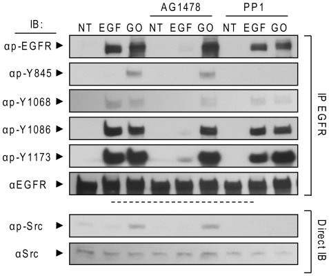
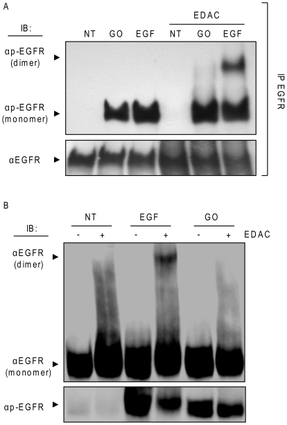
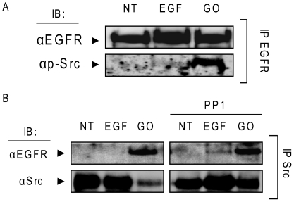
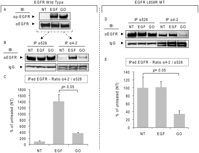
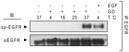
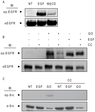
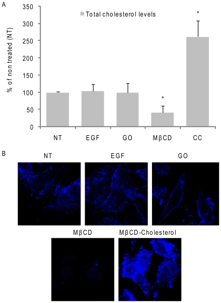
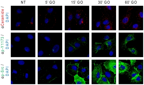
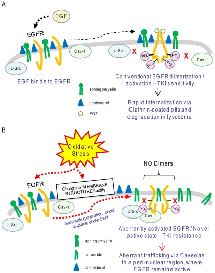
Crystallographic studies have offered understanding of how receptor tyrosine kinases from the ErbB family are regulated by their growth factor ligands. A conformational change of the EGFR (ErbB1) was shown to occur upon ligand binding, where a solely ligand-mediated mode of dimerization/activation was documented. However, this dogma of dimerization/activation was revolutionized by the discovery of constitutively active ligand-independent EGFR mutants. In addition, other ligand-independent activation mechanisms may occur. We have shown that oxidative stress (ox-stress), induced by hydrogen peroxide or cigarette smoke, activates EGFR differently than its ligand, EGF, thereby inducing aberrant phosphorylation and impaired trafficking and degradation of EGFR. Here we demonstrate that ox-stress activation of EGFR is ligand-independent, does not induce "classical" receptor dimerization and is not inhibited by the tyrosine kinase inhibitor AG1478. Thus, an unprecedented, apparently activated, state is found for EGFR under ox-stress. Furthermore, this activation mechanism is temperature-dependent, suggesting the simultaneous involvement of membrane structure. We propose that ceramide increase under ox-stress disrupts cholesterol-enriched rafts leading to EGFR re-localization into the rigid, ceramide-enriched rafts. This increase in ceramide also supports EGFR aberrant trafficking to a peri-nuclear region. Therefore, the EGFR unprecedented and activated conformation could be sustained by simultaneous alterations in membrane structure under ox-stress.


Conflict of interest statement

Competing Interests: The authors have declared that no competing interests exist.

Figures

Figure 1
Figure 1. H2O2 activation of EGFR is ligand-independent.
Serum-starved A549 cells were left intact or incubated with 40 nM monoclonal antibody 225 (mAb 225) for 1 hr on ice. Cells were then exposed for 15 min. at 37°C to 100 ng/ml EGF or 1 U/ml GO in the presence or absence of the mAb 225, as indicated. Immuno-precipitation (IP) of EGFR from cell lysates was performed using the mAb 528 and immuno-blotting (IB) for total (EGFR) and Tyr-phosphorylated EGFR (p-EGFR) was carried out as described in “Material and Methods”.
Figure 2
Figure 2. H2O2-induced EGFR phosphorylation is not inhibited by TKI AG1478 and is inhibited only at tyrosine 845 by Src family kinase inhibitor PP1.
Serum-starved A549 cells were incubated (or not) with 1 µM AG1478 or 5 µM PP1 for 30 min. Then, the cells were treated for 30 min. with 100 ng/ml EGF or 1 U/ml GO, as indicated. EGFR was IPed from cell lysates, resolved by SDS-PAGE and IBed for total receptor, total tyrosine phosphorylation (p-EGFR) and specific Tyr-residue phosphorylation level (Y845, Y1068, Y1086, and Y1173). Protein aliquots of the cell lysates were also directly IBed for total and Y416 phosphorylated (active) c-Src (p-Src).
Figure 3
Figure 3. H2O2-induced EGFR phosphorylation is inhibited by TKI AG1478 in crude membrane fractions.
A549 crude membrane fractions were prepared as indicated in “Material and Methods”. Membrane fractions were incubated with 1 µM AG1478 for 30 min. at 4°C and then incubated for 30 min. with 100 ng/ml EGF or 300 µM H2O2 at 37°C. A. Membrane proteins were separated by SDS-PAGE and IBed for total and Tyr-phosphorylated EGFR, as indicated. B. The histogram represents the levels of Tyr-phosphorylation of EGFR under different conditions, reported as fold-increase/decrease of the non treated (NT) sample; St-Deviations are indicated.
Figure 4
Figure 4. H2O2 activation of EGFR does not induce receptor dimerization.
A. Serum-starved A549 cells were exposed (or not) to 100 ng/ml EGF or 1 U/ml GO for 15 min. Then, the cross linking reagent EDAC (1 mM) was added and incubation was continued for an additional 15 min. EGFR was IPed from cell lysates, resolved by SDS-PAGE and IBed for total and Tyr-phosphorylated EGFR as indicated; presence of EGFR dimers is indicated. B. Serum-starved NIH-3T3 cells over-expressing wild type EGFR were treated as in A; 50 µg of total cell lysates were IBed for total and Tyr-phosphorylated EGFR as indicated.
Figure 5
Figure 5. EGFR directly interacts with c-Src under H2O2 and such interaction is not abolished by Src family kinase inhibitor PP1.
A549 cells were incubated (or not) with 5 µM PP1 for 45 min. and then treated (or not) for 15 min. with 100 ng/ml EGF or 30 min. 1 U/ml GO. A. EGFR was IPed from total cell lysates with the mAb 528 and IBed for Y416 phosphorylated c-Src (p-Src) and for total EGFR, as indicated. B. c-Src was IPed from total cell lysates and IBed for total c-Src and EGFR, as indicated.
Figure 6
Figure 6. Oxidative stress induces a novel active conformation of EGFR.
A. A549 cells were treated (or not) with 100 ng/ml EGF for 15 min. or with 1 U/ml GO for 30 min; Tyr-phosphorylation (p-Y20) and total EGFR levels were assessed by IB, as indicated. B. EGFR was IPed from 300 µg of the total cell lysates using either α528 or α4-2 Ab and then IBed with αEGFR Ab (2232). Samples IBs and the IgG heavy chains (stained with αmouse Ab) of the Abs used in the IPs are shown. The histogram in C represents the averaged ratio between total EGFR (IPed with α528 Ab) and the EGFR IPed with the α4-2 Ab (which has affinity for the “classical” EGF-induced active receptor conformation) of three independent experiments, quantified by densitometry of the bands; St-Dev are indicated. D. NIH-3T3 cells stably over-expressing L858R EGFR MT were treated as in A and EGFR was IPed as in B with either α528 or α4-2 Ab. Sample IBs of the IPs are shown for both EGFR (α2232) and IgG (αmouse Ab), as indicated. The graphic in E. represents the averaged ratio between total EGFR (IPed with α528 Ab) and EGFR IPed with the α4-2 Ab, quantified by densitometry of the bands (as in C); St-Dev are indicated.
Figure 7
Figure 7. H2O2 activation of EGFR is temperature-dependent.
Serum-starved A549 cells were left intact or incubated for 30 min. with 1 U/ml GO or 15 min. 100 ng/ml EGF at the indicated temperatures (T). EGFR was IPed from cell lysates and IBed for total and Tyr-phosphorylated EGFR, as indicated.
Figure 8
Figure 8. Cholesterol levels modulate EGFR activation by H2O2.
A. Serum-starved A549 cells were treated, or not, with 100 ng/ml EGF for 15 min. or 2% (w/v) MβCD for 1 h and cell lysates were IBed for total and Tyr-phosphorylated EGFR. B and C. Cells were treated with EGF as in A or with 1 U/ml GO for 30 min. in the absence or presence of 2 mM MβCD-cholesterol complex (CC), prepared as described in “Material and Methods”. Cell lysates were separated by SDS-PAGE and IBed for total and Tyr-phosphorylated EGFR (B) or total and Y416 phosphorylated Src (C).
Figure 9
Figure 9. H2O2-induced ox-stress does not deplete cellular cholesterol.
Serum-starved A549 cells were treated, or not, with 100 ng/ml EGF for 15 min. or 1 U/ml GO for 30 min. or 2% (w/v) MβCD for 1 h or 2 mM MβCD-cholesterol complexes (CC) for 30 min. A. Total cell cholesterol levels were measured after lipid extractions and normalized per protein unit, as described in “Material and Methods”; the values in the histogram are reported as % over non treated (NT) cells; St-Devs are indicated and “*” means p<0.05 in respect to control (NT); n = 3. B. Cells were fixed with paraformaldehyde (see “Material and Methods”) and stained for cholesterol using the sterol-binding probe filipin (50 µg/ml for 30 min.).
Figure 10
Figure 10. Ox-stress induces sustained increase of ceramide levels and activation of EGFR and c-Src.
A549 cells were seeded on cover-glasses, serum starved for 1 h and treated (or not) with 1 U/ml GO as indicated. After treatments, ceramide, EGFR phosphorylated on Y1173 and Y416 phosphorylated c-Src were localized in situ by IF as indicated in “Material and Methods”; nuclei were stained by DAPI.
Figure 11
Figure 11. Under H2O2-induced ox-stress elevated ceramide co-localizes with active EGFR and c-Src.
A549 cells were seeded on cover-glasses, serum starved for 1 h and treated (or not) with 1 U/ml GO as indicated. After treatments, ceramide (A and B), EGFR phosphorylated on Y1173 (A) and Y416 phosphorylated c-Src (B) were localized in situ by IF as indicated in “Material and Methods”; nuclei were stained by DAPI. White arrows indicate regions where p-Y1173 EGFR and ceramide (A) or p-Y416 c-Src (p-Src) and ceramide (B) co-localized under ox-stress (GO). Z-stack sections of cells have been discriminated by confocal microscopy: the panels show the merge of all of the Z-stack sections.
Figure 12
Figure 12. Proposed model of EGFR activation under ox-stress.
A. Conventional activation/dimerization of EGFR upon stimulation by the ligand EGF. B. Ox-stress could cause a change in membrane structure/fluidity by increasing cellular ceramide levels, which could affect cholesterol distribution. This, together with possible direct effect of ox-stress on EGFR, induces, or stabilizes, a novel acquired active conformation of EGFR that is bound by active c-Src and caveolin-1 (Cav-1). Such aberrantly active EGFR does not dimerize “conventionally” and becomes resistant to TKI drug, while it traffics via caveolae to an unidentified peri-nuclear region, where it remains active. Please note: Cav-1 binding to EGFR under ox-stress was demonstrated by our group before .

References

    1. Yarden Y, Sliwkowski MX. Untangling the ErbB signalling network. Nat Rev Mol Cell Biol. 2001;2:127–137. - PubMed
    1. Lu Z, Jiang G, Blume-Jensen P, Hunter T. Epidermal growth factor-induced tumor cell invasion and metastasis initiated by dephosphorylation and downregulation of focal adhesion kinase. Mol Cell Biol. 2001;21:4016–4031. - PMC - PubMed
    1. Schlessinger J. Ligand-induced, receptor-mediated dimerization and activation of EGF receptor. Cell. 2002;110:669–672. - PubMed
    1. Schlessinger J. The epidermal growth factor receptor as a multifunctional allosteric protein. Biochemistry. 1988;27:3119–3123. - PubMed
    1. Cochet C, Kashles O, Chambaz EM, Borrello I, King CR, et al. Demonstration of epidermal growth factor-induced receptor dimerization in living cells using a chemical covalent cross-linking agent. J Biol Chem. 1988;263:3290–3295. - PubMed

Publication types

MeSH terms