Evolution of cagA oncogene of Helicobacter pylori through recombination
- PMID: 21853141
- PMCID: PMC3154945
- DOI: 10.1371/journal.pone.0023499
Evolution of cagA oncogene of Helicobacter pylori through recombination
Abstract
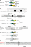
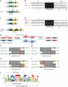
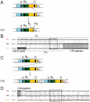
Helicobacter pylori is a gastric pathogen that infects half the human population and causes gastritis, ulcers, and cancer. The cagA gene product is a major virulence factor associated with gastric cancer. It is injected into epithelial cells, undergoes phosphorylation by host cell kinases, and perturbs host signaling pathways. CagA is known for its geographical, structural, and functional diversity in the C-terminal half, where an EPIYA host-interacting motif is repeated. The Western version of CagA carries the EPIYA segment types A, B, and C, while the East Asian CagA carries types A, B, and D and shows higher virulence. Many structural variants such as duplications and deletions are reported. In this study, we gained insight into the relationships of CagA variants through various modes of recombination, by analyzing all known cagA variants at the DNA sequence level with the single nucleotide resolution. Processes that occurred were: (i) homologous recombination between DNA sequences for CagA multimerization (CM) sequence; (ii) recombination between DNA sequences for the EPIYA motif; and (iii) recombination between short similar DNA sequences. The left half of the EPIYA-D segment characteristic of East Asian CagA was derived from Western type EPIYA, with Amerind type EPIYA as the intermediate, through rearrangements of specific sequences within the gene. Adaptive amino acid changes were detected in the variable region as well as in the conserved region at sites to which no specific function has yet been assigned. Each showed a unique evolutionary distribution. These results clarify recombination-mediated routes of cagA evolution and provide a solid basis for a deeper understanding of its function in pathogenesis.
Conflict of interest statement
Figures


Similar articles
-
Sequence Polymorphism and Intrinsic Structural Disorder as Related to Pathobiological Performance of the Helicobacter pylori CagA Oncoprotein.Toxins (Basel). 2017 Apr 13;9(4):136. doi: 10.3390/toxins9040136. Toxins (Basel). 2017. PMID: 28406453 Free PMC article. Review.
-
Characterization of East-Asian Helicobacter pylori encoding Western EPIYA-ABC CagA.J Microbiol. 2022 Feb;60(2):207-214. doi: 10.1007/s12275-022-1483-7. Epub 2021 Nov 10. J Microbiol. 2022. PMID: 34757586
-
c-Src and c-Abl kinases control hierarchic phosphorylation and function of the CagA effector protein in Western and East Asian Helicobacter pylori strains.J Clin Invest. 2012 Apr;122(4):1553-66. doi: 10.1172/JCI61143. Epub 2012 Mar 1. J Clin Invest. 2012. PMID: 22378042 Free PMC article.
-
Systematic analysis of phosphotyrosine antibodies recognizing single phosphorylated EPIYA-motifs in CagA of East Asian-type Helicobacter pylori strains.BMC Microbiol. 2016 Sep 2;16(1):201. doi: 10.1186/s12866-016-0820-6. BMC Microbiol. 2016. PMID: 27590005 Free PMC article.
-
Malignant Helicobacter pylori-Associated Diseases: Gastric Cancer and MALT Lymphoma.Adv Exp Med Biol. 2019;1149:135-149. doi: 10.1007/5584_2019_363. Adv Exp Med Biol. 2019. PMID: 31016622 Review.
Cited by
-
A specific A/T polymorphism in Western tyrosine phosphorylation B-motifs regulates Helicobacter pylori CagA epithelial cell interactions.PLoS Pathog. 2015 Feb 3;11(2):e1004621. doi: 10.1371/journal.ppat.1004621. eCollection 2015 Feb. PLoS Pathog. 2015. PMID: 25646814 Free PMC article.
-
Association between cagA and vacA genotypes and pathogenesis in a Helicobacter pylori infected population from South-eastern Sweden.BMC Microbiol. 2012 Jul 2;12:129. doi: 10.1186/1471-2180-12-129. BMC Microbiol. 2012. PMID: 22747681 Free PMC article.
-
Sequence Polymorphism and Intrinsic Structural Disorder as Related to Pathobiological Performance of the Helicobacter pylori CagA Oncoprotein.Toxins (Basel). 2017 Apr 13;9(4):136. doi: 10.3390/toxins9040136. Toxins (Basel). 2017. PMID: 28406453 Free PMC article. Review.
-
Elevated levels of adaption in Helicobacter pylori genomes from Japan; a link to higher incidences of gastric cancer?Evol Med Public Health. 2015 Mar 18;2015(1):88-105. doi: 10.1093/emph/eov005. Evol Med Public Health. 2015. PMID: 25788149 Free PMC article.
-
Phylogeographic evidence of cognate recognition site patterns and transformation efficiency differences in H. pylori: theory of strain dominance.BMC Microbiol. 2013 Sep 19;13:211. doi: 10.1186/1471-2180-13-211. BMC Microbiol. 2013. PMID: 24050390 Free PMC article.
References
-
- Suerbaum S, Michetti P. Helicobacter pylori infection. N Engl J Med. 2002;347:1175–1186. - PubMed
-
- Fischer W, Karnholz A, Jimenez-Soto LF, Haas R. Type IV secretion systems in Helicobacter pylori. In: Yamaoka Y, editor. Helicobacter pylori: molecular genetics and cellular biology. Caister Academic Press; 2008. 115
-
- Higashi H, Tsutsumi R, Muto S, Sugiyama T, Azuma T, et al. SHP-2 tyrosine phosphatase as an intracellular target of Helicobacter pylori CagA protein. Science. 2002;295:683–686. - PubMed
Publication types
MeSH terms
Substances
LinkOut - more resources
Full Text Sources

