. 2011;6(8):e23512.
doi: 10.1371/journal.pone.0023512. Epub 2011 Aug 10.

An atypical unfolded protein response in heat shocked cells

Affiliations

An atypical unfolded protein response in heat shocked cells

Lonneke Heldens et al. PLoS One. 2011.

Abstract

Background: The heat shock response (HSR) and the unfolded protein response (UPR) are both activated by proteotoxic stress, although in different compartments, and share cellular resources. How these resources are allocated when both responses are active is not known. Insight in possible crosstalk will help understanding the consequences of failure of these systems in (age-related) disease.

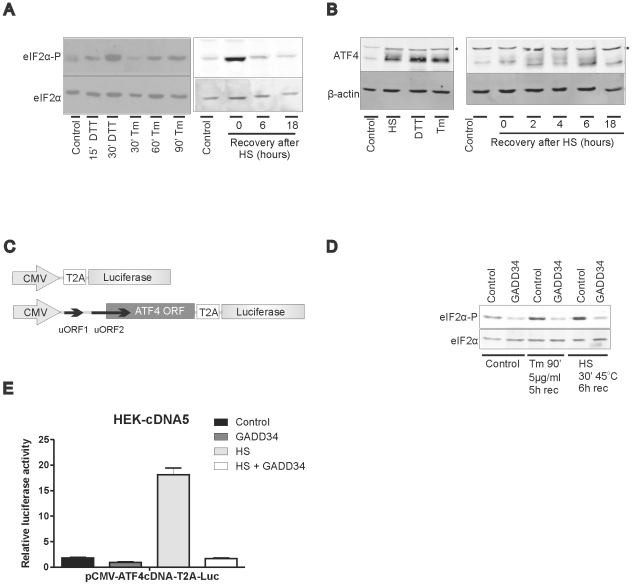
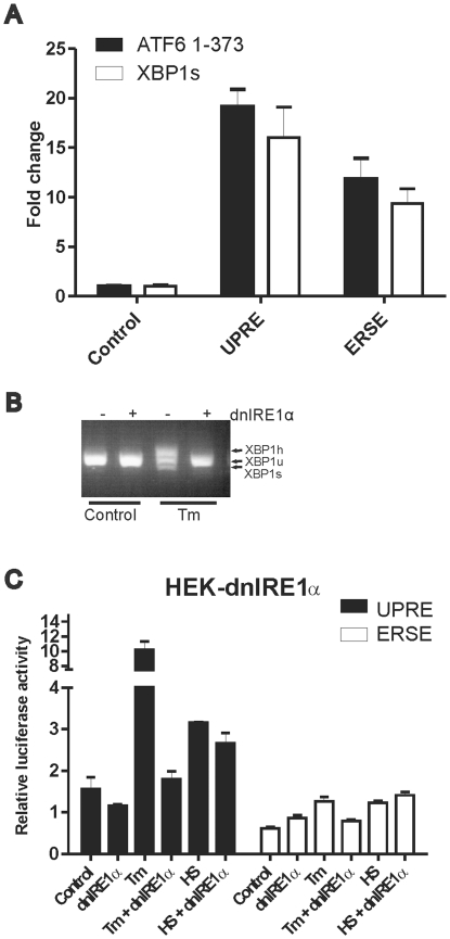
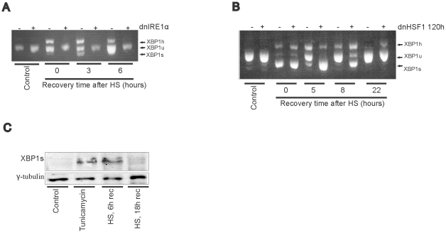
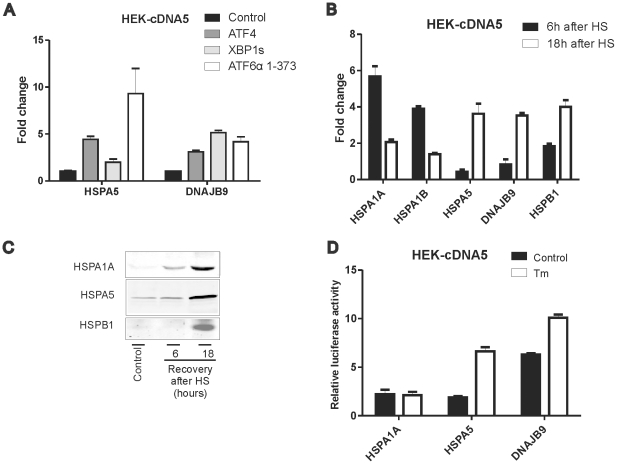
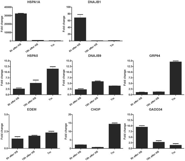
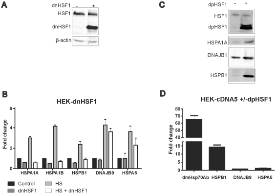
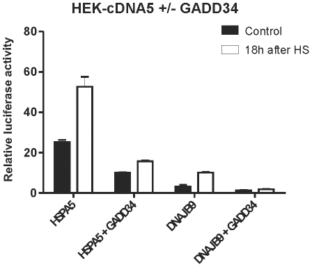
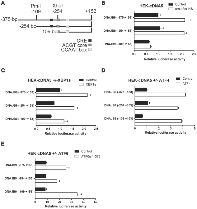
Results: In heat stressed HEK293 cells synthesis of the canonical UPR transcription factors XBP1s and ATF4 was detected as well as HSF1 independent activation of the promoters of the ER resident chaperones HSPA5 (BiP) and DNAJB9 (ERdj4). However, the heat stress activation of the DNAJB9 promoter, a XBP1s target, was not blocked in cells expressing a dominant negative IRE1α mutant, and thus did not require XBP1s. Furthermore, the DNA element required for heat stress activation of the DNAJB9 promoter is distinct from the ATF4 and ATF6 target elements; even though inhibition of eIF2α phosphorylation resulted in a decreased activation of the DNAJB9 promoter upon heat stress, suggesting a role for an eIF2α phosphorylation dependent product.

Conclusions: The initial step in the UPR, synthesis of transcription factors, is activated by heat stress but the second step, transcriptional transactivation by these factors, is blocked and these pathways of the UPR are thus not productive. Expression of canonical ER chaperones is part of the response of heat stressed cells but another set of transcription factors has been recruited to regulate expression of these ER chaperones.


Conflict of interest statement

Competing Interests: The authors have declared that no competing interests exist.

Figures

Figure 1
Figure 1. eIF2α-P dependent translation of the ATF4 ORF upon heat stress.
(A, B) eIF2α-P and ATF4 levels in ER or heat stressed cells. HEK-cDNA5 cells were treated with 10 mM DTT or 5 µg/ml tunicamycin for the indicated time to induce ER stress. Alternatively cells were exposed to a heat shock of 30′ at 45°C (HS) or left at 37°C (37°C). When heat shocked, cells were allowed to recover for the indicated time before harvesting. Cell lysates were subjected to SDS-PAGE and levels of phosphorylated eIF2α (eIF2α-P) were determined by western blotting with eIF2α as a loading control (panel A) or levels of ATF4 were determined by western blotting with β-actin as a loading control (panel B). The asterisk indicates a non specific band. (C) Schematic representation of the luciferase reporter constructs containing the ATF4 ORF. Translation of the luciferase code is dependent upon translation of the preceding ATF4 open reading frame. (D) Expression of C-term GADD34 decreases the level of eIF2α-P. HEK-cDNA5 cells were transfected with GADD34 or with an empty vector. Cell lysates of unstressed HEK-cDNA5 cells or HEK-cDNA5 cells exposed to tunicamycin or HS were subjected to SDS-PAGE and levels of phosphorylated eIF2α (eIF2α-P) were determined by western blotting. eIF2α was used as a loading control. (E) Translation of the ATF4 ORF in heat shocked cells is eIF2α-P dependent. HEK-cDNA5 cells were transfected with a mixture (4∶1∶5) of the indicated luciferase reporter, a βactin-βgal reporter, and the expression construct for C-term GADD34 or an empty vector. At 48 h after transfection, cells were exposed to a heat shock of 30′ at 45°C (HS) or left at 37°C (Control). When heat shocked, cells were allowed to recover for 7 hours and harvested. Harvested cells were assayed for reporter gene activities. The results are the average of three independent transfections (standard deviations are indicated by error bars).
Figure 2
Figure 2. UPRE and ERSE directed reporter gene activity.
(A) Effect of exogenous expression of ATF6 or spliced XBP1 on the activity of the UPRE-luciferase and ERSE-luciferase reporter constructs. HEK-cDNA5 cells were transfected with a mixture (4∶1∶5) of the indicated luciferase reporter, a βactin-βgal reporter, and pcDNA3.1-ATF6α (1–373), pcDNA5-XBP1s or an empty vector pcDNA5/FRT/TO. At 48 hours after transfection, cells were harvested and assayed for reporter gene activities. (B) dnIRE1α blocks XBP1 splicing. HEK-dnIRE1α cells were cultured in the absence or presence of doxycycline. Cells were treated with 10 µg/ml tunicamycin for 90′ to induce XBP1 splicing. Total RNA samples were analyzed by RT-PCR. PCR products represent unspliced (XBP1u), spliced (XBP1s) and a hybrid of spliced and unspliced XBP1 PCR products (XBP1h; see also Materials and methods). (C) HEK-cDNA5 and HEK-dnIRE1α cells were transfected with a mixture (9∶1) of the indicated luciferase reporter and a βactin-βgal reporter. At 24 hours after transfection dnIRE1α expression was induced by adding doxycycline. At 48 hours after transfection, cells were exposed to a heat shock of 30′ at 45°C (HS) or left at 37°C (37°C). When heat shocked, cells were allowed to recover for 18 hours and harvested. To induce ER stress, cells were exposed to 2 µg/ml tunicamycin for 24 hours. Harvested cells were assayed for reporter gene activities.
Figure 3
Figure 3. XBP1 splicing in heat shocked cells.
(A) HEK-dnIRE1α cells were cultured in the absence or presence of doxycycline. Cells were exposed to a heat shock of 30′ at 45°C (HS) or left at 37°C (no stress). When heat shocked, cells were allowed to recover for the indicated periods at 37°C. Total RNA samples were analyzed by RT-PCR. (B) HEK-dnHSF1 cells were cultured in the absence or presence of doxycycline for the indicated time. Cells were exposed to a heat shock (30′, 45°C), harvested at the indicated time point after heat shock, and subjected RT-PCR analysis to investigate the effect of HEK-dnHSF1 on XBP1 splicing after heat stress. (C) XBP1s levels in heat stressed cells. HEK-cDNA5 cells were exposed to a heat shock of 30′ at 45°C or left at 37°C. When heat shocked, cells were allowed to recover for the indicated time before harvesting. To induce ER stress, cells were treated with 5 µg/ml tunicamycin for 90 minutes. After 5 hours recovery cells were harvested. Cell lysates were subjected to SDS-PAGE and levels of XBP1s were determined by western blotting with γ-tubulin as a loading control.
Figure 4
Figure 4. Stress response of various promoters.
(A) ATF4, ATF6 and XBP1s activate the HSPA5 and DNAJB9 promoters. HEK-cDNA5 cells were transfected with a mixture (4∶1∶5) of the indicated luciferase reporter, a β-actin-βgal reporter, and pcDNA5-XBP1s, pcDNA5-ATF4ORF, pcDNA3.1-ATF6α (1–373) or the empty vector pcDNA5/FRT/TO. At 24 hours after transfection doxycyclin was added to induce expression of XBP1s or ATF4. At 48 hours after transfection, cells were harvested and assayed for reporter gene activities. For the levels of exogenously expressed XBP1 and ATF4, see Fig. S1. (B) Heat shock response of various promoters. HEK-cDNA5 cells were transfected with the indicated promoter reporter construct and a β-actin-β-galactosidase reporter (9∶1 ratio). At 48 h after transfection, cells were exposed to a heat shock of 30′ at 45°C or left at 37°C. When heat shocked, cells were allowed to recover for 6 hours or 18 hours, and assayed for reporter gene activities. The luciferase activity of the promoter constructs is relative to the activity of pGL3 control. (C) HEK-cDNA5 cells were exposed to a heat shock of 30′ at 45°C or left at 37°C. When heat shocked, cells were allowed to recover for the indicated time before harvesting. Cell lysates were subjected to SDS-PAGE and levels of HSPA1A, HSPA5 and HSPB1 were determined by western blotting. (D) ER stress activation of various promoters. HEK-cDNA5 cells were transfected with the indicated promoter reporter construct and a β-actin-β-galactosidase reporter (9∶1 ratio). At 48 h after transfection, cells were exposed to 2 µg/ml tunicamycin for 18 hours, and assayed for reporter gene activities. The luciferase activity of the promoter constructs is relative to the activity of pGL3 control.
Figure 5
Figure 5. Relative changes in transcript levels of ER responsive genes in heat shocked and tunicamycin treated cells.
HEK-cDNA5 cells were treated with 2 µg/ml tunicamycin for 24 hours to induce ER stress. Alternatively cells were exposed to a heat shock of 30′ at 45°C (HS) or left at 37°C (37°C). When heat shocked, cells were allowed to recover for the indicated time before harvesting. Total RNA was isolated and transcript levels were measured by QPCR. Fold induction of mRNA levels is plotted relative to GAPDH mRNA levels.
Figure 6
Figure 6. HSF1 dependency of various promoters.
(A) Expression of dnHSF1. HEK-dnHSF1 were cultured in the absence or presence of doxycycline to induce expression of dnHSF1. After harvesting the cells were subjected to SDS-PAGE and levels of endogeneous HSF1 and dnHSF1 were determined by western blotting with β-actin as a loading control. (B) The effects of dnHSF1 on basal and heat shock induced activity of various promoters. Cells were transfected with a mixture of the indicated luciferase reporter and a β -actin-β-galactosidase reporter (9∶1 ratio). At 24 hours after transfection doxycyclin was added to induce expression of dnHSF1. At 48 hours after transfection, cells were exposed to a heat shock of 30′ at 45°C or left at 37°C. When heat shocked, cells were allowed to recover for the indicated periods at 37°C. Harvested cells were assayed for reporter gene activities. (C) Expression of dpHSF1 leads to increased HSP levels. HEK-cDNA5 cells were tranfected with pcDNA5-dpHSF1. At 24 hours after transfection cells were cultured in the absence or presence of doxycycline to induce expression of dpHSF1. At 48 hours after transfection cells were harvested and subjected to SDS-PAGE and levels of endogenous HSF1, dpHSF1, HSPA1A, DNAJB1 and HSPB1 were determined by western blotting. (D) The effect of dpHSF1 on the activity of various promoters. HEK-cDNA5 cells were transfected with a mixture (4∶1∶5) of the indicated luciferase reporter, a βactin-βgal reporter, and pcDNA5-dpHSF1. At 24 hours after transfection doxycycline was added to induce expression of dpHSF1. At 48 hours after transfection, cells were harvested and assayed for reporter gene activities.
Figure 7
Figure 7. DNAJB9 promoter activity is not regulated by XBP1s in heat shocked cells.
(A) Activity of the DNAJB9 promoter in HEK-dnIRE1α cells. HEK-dnIRE1α cells were transfected with a mixture of the indicated luciferase reporter and a β -actin-β-galactosidase reporter (9∶1 ratio). At 24 hours after transfection doxycyclin was added to induce expression of dnIRE1α. At 48 hours after transfection, cells were exposed to a heat shock of 30′ at 45°C or left at 37°C. When heat shocked, cells were allowed to recover for 18 hours and harvested. Alternatively, cells were exposed to 2 µg/ml Tunicamycin for 18 hours. Harvested cells were assayed for reporter gene activities. (B) The DNAJB9 promoter can be activated by exogenous XBP1s. HEK-cDNA5 cells were transfected with a mixture (4∶1∶5) of the indicated luciferase reporter, a βactin-βgal reporter, and pcDNA5-XBP1s or the empty vector pcDNA5/FRT/TO. At 24 hours after transfection doxycyclin was added to induce expression of XBP1s. At 48 hours after transfection, cells were exposed to a heat shock of 30′ at 45°C or left at 37°C. When heat shocked, cells were allowed to recover for 18 hours, harvested and assayed for reporter gene activities.
Figure 8
Figure 8. eIF2α-P dependent DNAJB9 and HSPA5 promoter activity.
Activity of DNAJB9 and HSPA5 promoter in heat shocked cells is eIF2α-P dependent. HEK-cDNA5 cells were transfected with a mixture (4∶1∶5) of the indicated luciferase reporter, a βactin-βgal reporter, and the expression construct for GADD34 or an empty vector. At 48 hours after transfection, cells were exposed to a heat shock of 30′ at 45°C (HS) or left at 37°C (37°C). When heat shocked, cells were allowed to recover for 18 hours and harvested. Harvested cells were assayed for reporter gene activities.
Figure 9
Figure 9. DNAJB9 promoter deletion constructs.
(A) Schematic representation of the DNAJB9 promoter region. (B) Heat shock inducibility of DNAJB9 deletion constructs. HEK-pcDNA5 cells were transfected with a mixture of the indicated luciferase reporter and a βactin-β-galactosidase reporter (9∶1 ratio). At 48 hours after transfection, cells were exposed to a heat shock of 30′ at 45°C or left at 37°C. When heat shocked, cells were allowed to recover for 18 hours, harvested and assayed for reporter gene activities. (C,D,E) Effect of exogenous XBP1s, ATF4 or ATF6 expression on the activity of promoter deletion constructs. HEK-cDNA5 cells were transfected with a mixture (4∶1∶5) of the indicated luciferase reporter, a βactin-βgal reporter, and an expression construct as indicated or the empty vector pcDNA5/FRT/TO. At 24 hours after transfection doxcycyclin was added to induce XBP1s (C) or ATF4 expression (D). At 48 hours after transfection, cells were harvested and assayed for reporter gene activities.

Similar articles

Cited by

References

    1. Zhang K, Kaufman RJ. The unfolded protein response: a stress signaling pathway critical for health and disease. Neurology. 2006;66:S102–109. - PubMed
    1. Balch WE, Morimoto RI, Dillin A, Kelly JW. Adapting proteostasis for disease intervention. Science. 2008;319:916–919. - PubMed
    1. Powers ET, Morimoto RI, Dillin A, Kelly JW, Balch WE. Biological and chemical approaches to diseases of proteostasis deficiency. Annu Rev Biochem. 2009;78:959–991. - PubMed
    1. Broadley SA, Hartl FU. The role of molecular chaperones in human misfolding diseases. FEBS Lett. 2009;583:2647–2653. - PubMed
    1. Morimoto RI. Regulation of the heat shock transcriptional response: cross talk between a family of heat shock factors, molecular chaperones, and negative regulators. Genes Dev. 1998;12:3788–3796. - PubMed

Publication types

MeSH terms