. 2012 Jan;61(1):101-8.
doi: 10.1007/s00262-011-1094-3. Epub 2011 Aug 19.

Upregulation of B7-H1 expression is associated with macrophage infiltration in hepatocellular carcinomas

Affiliations

Upregulation of B7-H1 expression is associated with macrophage infiltration in hepatocellular carcinomas

Jie Chen et al. Cancer Immunol Immunother. 2012 Jan.

Abstract

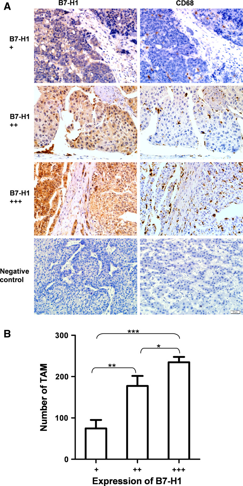
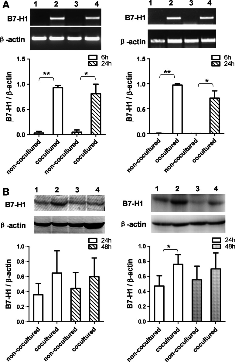
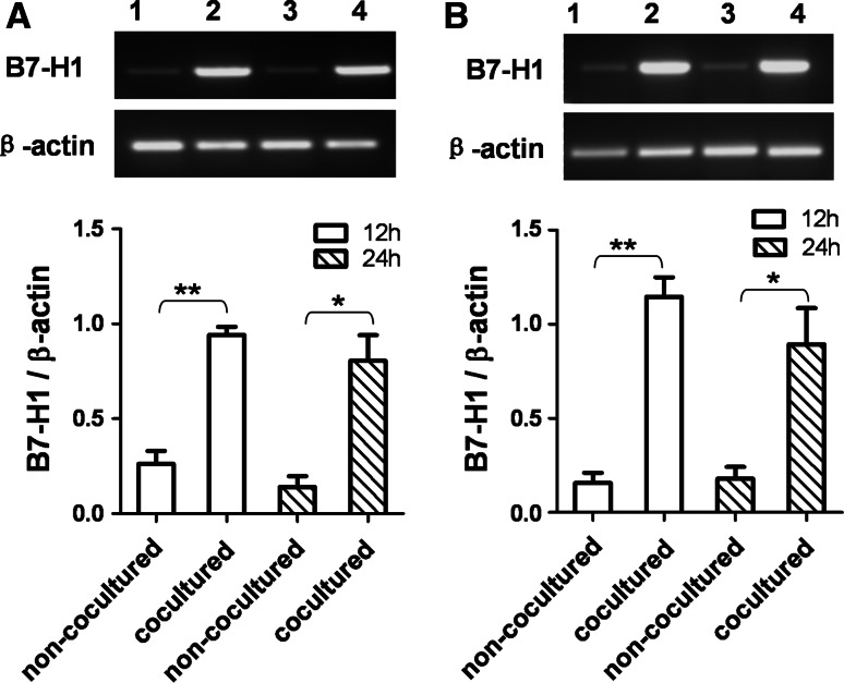
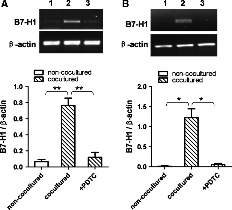
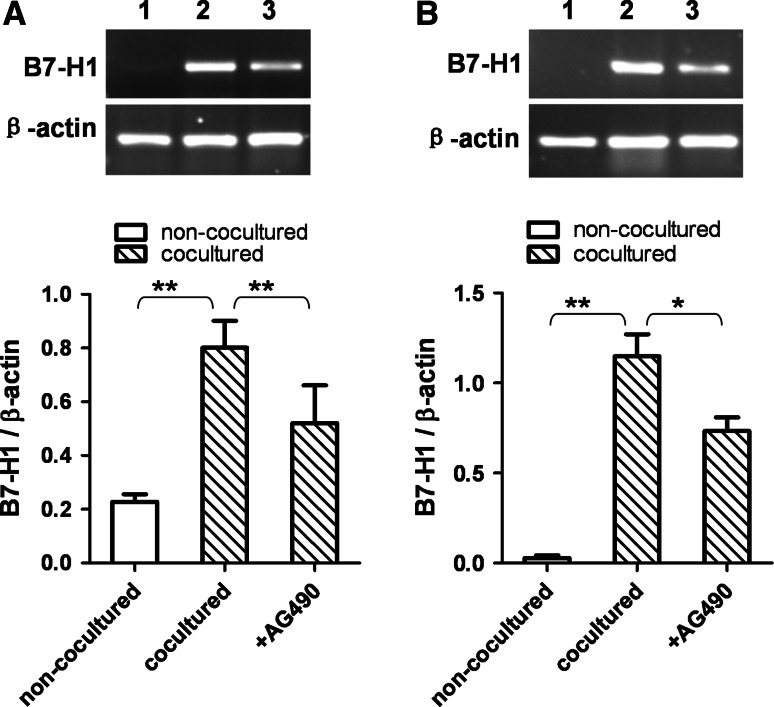
The overexpression of B7-H1 in hepatocellular carcinoma (HCC) mediates HCC immune escape and obstructs the immunotherapy based on tumor-specific CD8+ T cells. Tumor-associated macrophages (TAM) are a major component of cancer-related inflammation and play a central role in tumor promotion. To classify the mechanism underlying the overexpression of B7-H1 in HCC, we examined B7-H1 expression and TAM infiltration in 63 cases of human HCC samples using immunohistochemistry method and found that B7-H1 overexpression was associated with TAM infiltration in HCC tissues. Furthermore, B7-H1 expression was upregulated at both mRNA level and protein level in HCC cells (BEL-7402 and SMMC-7721) cocultured with macrophages in a transwell system. The upregulation of B7-H1 expression induced by macrophage was inhibited by blocking NF-κB or STAT3 signal pathways. These results suggest that overexpression of B7-H1 in HCC may be induced by inflammatory microenvironment involving macrophages and imply that anti-inflammation therapy might be preventive for immune escape and assistant for immunotherapy of HCC.


Figures

Fig. 1
Fig. 1
B7-H1 overexpression was associated with TAM infiltration in HCC tissues. a Expression of B7-H1 and CD68 was tested in consecutive sections from 63 HCC patients using immunohistochemitry method. CD68-positive staining indicated the TAM infiltration. The strength of B7-H1 expression was classified into grade +, grade ++ and grade +++ using Image-Pro Plus 6.0 software. Three representative cases showed B7-H1 expression and TAM infiltration. Positive cells were stained brown (×200). Negative controls were performed by omitting the primary antibodies. b Statistical analysis of the relationship between B7-H1 expression and TAM infiltration. TAM number in HCC tissues with high expression of B7-H1 was significantly higher than that in HCC tissues with low expression of B7-H1. Data shown are expressed as means ± SE. Asterisk indicates P < 0.05. Double asterisk indicates P < 0.01. Triple asterisk indicates P < 0.001
Fig. 2
Fig. 2
B7-H1 expression was upregulated in HCC cells by PMA-treated THP-1 cells. a B7-H1 mRNA expression was upregulated in HCC cells by PMA-treated THP-1 cells. After coculture for 6 and 24 h with PMA-treated THP-1 cells in a transwell system, BEL-7402 (left figure) and SMMC-7721 (right figure) cells were harvested, and B7-H1 mRNA expression was determined by RT-PCR. Lane 1, non-cocultured 6 h; lane 2, cocultured 6 h; lane 3, non-cocultured 24 h; lane 4, cocultured 24 h. Densitometric data shown are means ± SE of the results for three independent experiments. b B7-H1 protein expression was upregulated in HCC cells by PMA-treated THP-1 cells. HCC cells were cocultured with PMA-treated THP-1 cells in a transwell system. After 24 and 48 h of coculture, BEL-7402 (left figure) and SMMC-7721 (right figure) cells were harvested, and B7-H1 protein expression was determined by Western blot. Lane 1, non-cocultured 24 h; lane 2, cocultured 24 h; lane 3, non-cocultured 48 h; lane 4, cocultured 48 h. Densitometric data shown are means ± S.E of the results for three independent experiments. Asterisk indicates P < 0.05. Double asterisk indicates P < 0.01
Fig. 3
Fig. 3
B7-H1 expression was upregulated in HCC cells by PMA-induced human macrophages After coculture for 12 and 24 h with PMA-induced human macrophages in a transwell system, BEL-7402 (a) and SMMC-7721 (b) cells were harvested, and B7-H1 mRNA expression was determined by RT-PCR. Lane 1, non-cocultured 12 h; lane 2, cocultured 12 h; lane 3, non-cocultured 24 h; lane 4, cocultured 24 h. Densitometric data shown are means ± S.E of the results for three independent experiments. Asterisk indicates P < 0.05. Double asterisk indicates P < 0.01
Fig. 4
Fig. 4
The induction of B7-H1 expression was inhibited by blocking NF-κB pathway. BEL-7402 and SMMC-7721 cells were treated with PDTC (10 ng/mL) for one h before coculture with PMA-treated THP-1 cells. After 6 h of coculture, BEL-7402 (a) and SMMC-7721 (b) cells were harvested, and B7-H1 mRNA expression was determined by RT-PCR. Lane 1, non-cocultured; lane 2, cocultured; lane 3, PDTC-treated and cocultured. Densitometric data shown are the means ± SE of triplicate experiments. Asterisk indicates P < 0.05. Double asterisk indicates P < 0.01
Fig. 5
Fig. 5
The induction of B7-H1 expression was inhibited by blocking STAT3 pathway. BEL-7402 and SMMC-7721 cells were treated with AG490 (20 ng/mL) for 24 h before coculture with PMA-treated THP-1 cells. After 6 h of coculture, BEL-7402 (a) and SMMC-7721 (b) cells were harvested, and B7-H1 mRNA expression was determined by RT-PCR. Lane 1, non-cocultured; lane 2, cocultured; lane 3, AG490-treated and cocultured. Densitometric data shown are the means ± SE of triplicate experiments. Asterisk indicates P < 0.05. Double asterisk indicates P < 0.01

Similar articles

Cited by

References

    1. Andreana L, Burroughs AK. Treatment of early hepatocellular carcinoma: how to predict and prevent recurrence. Dig Liver Dis. 2010;42(Suppl 3):S249–S257. doi: 10.1016/S1590-8658(10)60513-0. - DOI - PubMed
    1. Nowak AK, Chow PK, Findlay M. Systemic therapy for advanced hepatocellular carcinoma: a review. Eur J Cancer. 2004;40(10):1474–1484. doi: 10.1016/j.ejca.2004.02.027. - DOI - PubMed
    1. Hiroishi K, Eguchi J, Baba T, Shimazaki T, Ishii S, Hiraide A, Sakaki M, Doi H, Uozumi S, Omori R, Matsumura T, Yanagawa T, Ito T, Imawari M. Strong CD8(+) T-cell responses against tumor-associated antigens prolong the recurrence-free interval after tumor treatment in patients with hepatocellular carcinoma. J Gastroenterol. 2010;45(4):451–458. doi: 10.1007/s00535-009-0155-2. - DOI - PubMed
    1. Sun JC, Pan K, Chen MS, Wang QJ, Wang H, Ma HQ, Li YQ, Liang XT, Li JJ, Zhao JJ, Chen YB, Pang XH, Liu WL, Cao Y, Guan XY, Lian QZ, Xia JC. Dendritic cells-mediated CTLs targeting hepatocellular carcinoma stem cells. Cancer Biol Ther. 2010;10(4):368–375. doi: 10.4161/cbt.10.4.12440. - DOI - PubMed
    1. Zhang HH, Mei MH, Fei R, Liao WJ, Wang XY, Qin LL, Wang JH, Wei L, Chen HS. Regulatory T cell depletion enhances tumor specific CD8 T-cell responses, elicited by tumor antigen NY-ESO-1b in hepatocellular carcinoma patients, in vitro. Int J Oncol. 2010;36(4):841–848. - PubMed

Publication types

MeSH terms