Controlled Clinical Trial
. 2011 Aug 15;52(4):488-96.
doi: 10.3325/cmj.2011.52.488.

Clinical sensitivity and specificity of multiple T2-hyperintensities on brain magnetic resonance imaging in diagnosis of neurofibromatosis type 1 in children: diagnostic accuracy study

Affiliations
Controlled Clinical Trial

Clinical sensitivity and specificity of multiple T2-hyperintensities on brain magnetic resonance imaging in diagnosis of neurofibromatosis type 1 in children: diagnostic accuracy study

Zlatko Sabol et al. Croat Med J. .

Abstract

Aim: To determine the prevalence, number, and location of multiple (≥2) T2-hyperintensities on brain magnetic resonance imaging (MRI) in children with neurofibromatosis type 1 (NF1) and their correlation with age, and to establish their sensitivity, specificity, and accuracy for the diagnosis of NF1 in children, especially in the early age (2-7 years).

Methods: We performed a cross-sectional study of 162 patients with NF1 from Croatian Neurofibromatosis Association Database and 163 control children between the ages of 2 and 18 years who underwent brain MRI between 1989 and 2009.

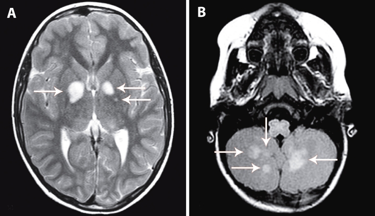
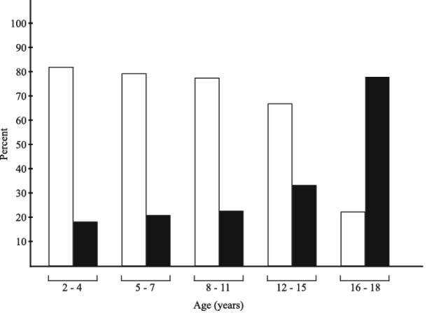
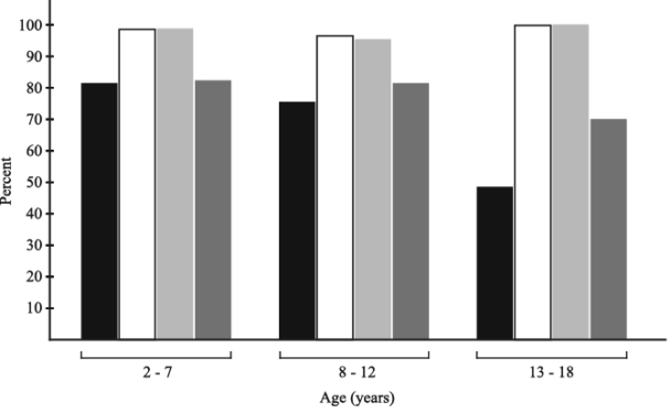
Results: Multiple T2-hyperintensities were present in 74% of NF1 patients and 1.8% of controls. They were mainly located in the basal ganglia, brainstem, and cerebellum and were significantly decreased in prevalence and number in the older age. T2-hyperintensities had excellent diagnostic accuracy with the area under the receiver operating characteristic (ROC) curve of 0.849 and 95% confidence interval (CI) of 0.805-0.886. The diagnostic sensitivity, specificity, and accuracy rate of T2-hyperintensities for NF1 were highest in the youngest age (2-7 years): 81% (95% CI 71%-89.1%), 99% (95% CI 92.3%-100%), and 85.8 (95% CI 83.3-93.8), respectively.

Conclusion: This study strongly suggests the inclusion of T2-hyperintensities on brain MRI on the list of diagnostic criteria for NF1, especially in children of early age, when the clinical penetration of the NF1 gene has not yet been completely finished.


Figures

Figure 1
Figure 1
Multiple hyperintensities on T2-weighted cranial magnetic resonance imaging in children with neurofibromatosis type 1. Axial image shows multiple oval discrete lesions with increased intensity (arrows) in the globus pallidus (A); axial fluid attenuated inversion recovery image shows multiple diffuse T2-hyperintensities (arrows) in the cerebellum (B).
Figure 2
Figure 2
Prevalence of T2-hyperintensities on brain magnetic resonance imaging (MRI) in different age groups of children with neurofibromatosis type 1. The frequency of T2-hyperintensities significantly decreased from age 2-4 years to age 16-18 years (χ2 = 12.03, P = 0.002). Open bars – T2-hyperintensities present; closed bars – T2-hyperintensities absent.
Figure 3
Figure 3
The correlation of the number of T2-hyperintensities and age in 119 children with neurofibromatosis type 1. Pearson correlation showed a significant correlation between the number T2-hyperintensities and age of patients with NF1 (r2 linear = 0.100, P = 0.011).
Figure 4
Figure 4
Clinical diagnostic sensitivity, specificity, and positive and negative predictive value of T2-hyperintensities in patients with neurofibromatosis type 1 of different age. Closed bars – sensitivity; open bars – specificity; light gray – positive predictive value; dark gray – negative predictive value.
Figure 5
Figure 5
Receiver operating characteristic (ROC) curve of T2-hyperintensities on brain magnetic resonance imaging. ROC curve (full line) and confidence interval (dashed line) for T2-hyperintensities. Gray line defines the area of 0.5.

Similar articles

Cited by

References

    1. Huson SM, Harper PS, Compston DAS. Von Recklinghausen neurofibromatosis. A clinical and population study in South East Wales. Brain. 1988;111:1355–81. doi: 10.1093/brain/111.6.1355. - DOI - PubMed
    1. Barker D, Wright E, Nguyen K, Cannon L, Fain P, Goldgar D, et al. Gene for von Recklinghausen neurofibromatosis is in pericentromeric region of chromosome 17. Science. 1987;236:1100–2. doi: 10.1126/science.3107130. - DOI - PubMed
    1. Viskochil D, Cawthon R, O'Connel P, Xu GF, Stevens J, Culver M, et al. The gene encoding the oligodendrocyte-myelin glycoprotein is embedded within the neurofibromatosis type 1 gene. Mol Cell Biol. 1991;11:906–12. - PMC - PubMed
    1. Marchuk DA, Saulino AM, Tavakkol RL, Swaroop M, Wallace MR, Andersen LB, et al. cDNA cloning of the type 1 neurofibromatosis gene: complete sequence of the NF1 gene product. Genomics. 1991;11:931–40. doi: 10.1016/0888-7543(91)90017-9. - DOI - PubMed
    1. Crowe FW, Schull WJ, Neel JV. A clinical, pathological, and genetic study of multiple neurofibromatosis. Springfield, (IL): Charles C. Thomas; 1956.

Publication types