. 2011 Nov;176(5):636-48.
doi: 10.1667/rr2422.1. Epub 2011 Aug 19.

Laminin 332 deposition is diminished in irradiated skin in an animal model of combined radiation and wound skin injury

Affiliations

Laminin 332 deposition is diminished in irradiated skin in an animal model of combined radiation and wound skin injury

M M Jourdan et al. Radiat Res. 2011 Nov.

Abstract

Skin exposure to ionizing radiation affects the normal wound healing process and greatly impacts the prognosis of affected individuals. We investigated the effect of ionizing radiation on wound healing in a rat model of combined radiation and wound skin injury. Using a soft X-ray beam, a single dose of ionizing radiation (10-40 Gy) was delivered to the skin without significant exposure to internal organs. At 1 h postirradiation, two skin wounds were made on the back of each rat. Control and experimental animals were euthanized at 3, 7, 14, 21 and 30 days postirradiation. The wound areas were measured, and tissue samples were evaluated for laminin 332 and matrix metalloproteinase (MMP) 2 expression. Our results clearly demonstrate that radiation exposure significantly delayed wound healing in a dose-related manner. Evaluation of irradiated and wounded skin showed decreased deposition of laminin 332 protein in the epidermal basement membrane together with an elevated expression of all three laminin 332 genes within 3 days postirradiation. The elevated laminin 332 gene expression was paralleled by an elevated gene and protein expression of MMP2, suggesting that the reduced amount of laminin 332 in irradiated skin is due to an imbalance between laminin 332 secretion and its accelerated processing by elevated tissue metalloproteinases. Western blot analysis of cultured rat keratinocytes showed decreased laminin 332 deposition by irradiated cells, and incubation of irradiated keratinocytes with MMP inhibitor significantly increased the amount of deposited laminin 332. Furthermore, irradiated keratinocytes exhibited a longer time to close an artificial wound, and this delay was partially corrected by seeding keratinocytes on laminin 332-coated plates. These data strongly suggest that laminin 332 deposition is inhibited by ionizing radiation and, in combination with slower keratinocyte migration, can contribute to the delayed wound healing of irradiated skin.


Figures

FIG. 1
FIG. 1
Time course of radiation and wound skin injury in rats irradiated with a single dose of radiation. The rows depict clinical phenotype at day 7, 14 and 30 postirradiation, while columns represent the different radiation doses.
FIG. 2
FIG. 2
Time course of skin injury score after single doses of radiation. Each point represents data from 7 animals. The lines trace the median values; the bars show lowest and highest score at the particular time. All scores beyond 3 days are significantly elevated (P < 0.001 compared to age-matched normal animals).
FIG. 3
FIG. 3
Time course of wound closure after single doses of radiation. Each point represents data from 7 animals. The columns represent mean values, bars ± SEM, and asterisks statistically significant decreases in wound closure (P < 0.05 compared to unirradiated animals).
FIG. 4
FIG. 4
Time course and dose dependence of laminin 332 protein staining in irradiated and sham-irradiated skin. Panel A: The upper row shows laminin 332 staining at day 14, the lower row at day 30. The arrows point toward laminin 332-specific staining in the epidermal and hair follicle basement membranes. All images were taken at 20× magnification. The intensity of laminin 332 staining was quantified at (panel B) 14 and (panel C) 30 days postirradiation using the NIH image J program 1.43; asterisks indicate statistically significant decreases in staining (P < 0.001 compared to unirradiated animals).
FIG. 5
FIG. 5
Time course of laminin 332 protein staining in irradiated and sham-irradiated wounds. Panel A: The upper row shows laminin 332 staining at day 3, 7 and 14 in sham-irradiated wounds. The lower row shows laminin 332 staining at the same time in wounds from animals irradiated with 30 Gy. The arrows point toward laminin 332 deposition at the edge of the wound as well as in the provisional wound bed. All images were taken at 20× magnification. Panel B: The intensity of laminin 332 staining was quantified in the wounds at 3, 7 and 14 days postirradiation by NIH image J program 1.43; asterisks indicate statistically significant decreases in staining (P < 0.001 compared to unirradiated animals).
FIG. 6
FIG. 6
Immunoelectron microscopy evaluation of laminin 332 deposition in skin 120 days postirradiation. Panel A shows immunogold particles bound to laminin 332 in irradiated skin (12.5 Gy), panel B in unirradiated skin. Stars indicate basal keratinocytes, closed circles indicate collagen bundles, and arrows point to immunogold particles.
FIG. 7
FIG. 7
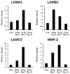
Time-course assessment of LAMA3, LAMB3, LAMC2 and MMP2 mRNA expression by qRT-PCR from skin samples taken from control and irradiated (30 Gy) wound edge. Each point represents data from RNA samples pooled from 5 animals. The data were normalized to the levels of the housekeeping gene GAPDH in sham-irradiated wounded skin and were expressed as the relative change.
FIG. 8
FIG. 8
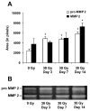
Time-course assessment of MMP2 activity by gelatin zymography. Panel A: Densitometric evaluation of pro MMP2 and MMP2 at day 3, 4 and 14 in wound skin samples from sham-irradiated animals and animals irradiated with 30 Gy. Each point represents data from 4 animals; asterisks indicate statistically significant increased activity (P < 0.001 compared to unirradiated animals). Panel B: A representative gel demonstrating pro-MMP2 (72 kDa) and MMP2 (62 kDa) bands in irradiated and sham-irradiated wounds.
FIG. 9
FIG. 9
Western blot analysis of laminin 332 deposition and processing in nonirradiated and irradiated cultured rat keratinocytes. Panel A: Representative picture of laminin 332 extracted from keratinocytes irradiated with 0 or 2 Gy and incubated in the presence or absence of the MMP inhibitor (MMPi) 1,10-phenanthroline. Panel B: Densitometric evaluation of all laminin 332 subunits 48 h postirradiation. n = 3; error bars are standard deviations.
FIG. 10
FIG. 10
In vitro keratinocyte migration assay. Panel A: Representative pictures of rat keratinocytes irradiated with 0, 2 or 10 Gy and scratch wounded immediately after irradiation. Pictures were taken 48 h after irradiation. Panel B: Evaluation of the scratch width of irradiated and sham-irradiated keratinocytes using the NIH Image J program 1.43. n = 6, error bars are standard deviations. Asterisks label statistically significant delayed width closure (2 Gy, P = 0.008; 10 Gy, P = 0.001) compared to nonirradiated animals.

References

    1. Messerschmidt O. Whole-body irradiation plus skin wound: animal experiments on combined injuries. Br J Radiol Suppl. 1986;19:64–7. - PubMed
    1. DiCarlo AL, Hatchett RJ, Kaminski JM, Ledney GD, Pellmar TC, Okunieff P, et al. Medical countermeasures for radiation combined injury: radiation with burn, blast, trauma and/or sepsis. report of an NIAID Workshop, March 26–27, 2007. Radiat Res. 2008;169:712–21. - PMC - PubMed
    1. Augustine AD, Gondre-Lewis T, McBride W, Miller L, Pellmar TC, Rockwell S. Animal models for radiation injury, protection and therapy. Radiat Res. 2005;164:100–9. - PubMed
    1. Ran X, Cheng T, Shi C, Xu H, Qu J, Yan G, et al. The effects of total-body irradiation on the survival and skin wound healing of rats with combined radiation-wound injury. J Trauma. 2004;57:1087–93. - PubMed
    1. Kiang JG, Jiao W, Cary LH, Mog SR, Elliott TB, Pellmar TC, et al. Wound trauma increases radiation-induced mortality by activation of iNOS pathway and elevation of cytokine concentrations and bacterial infection. Radiat Res. 2010;173:319–32. - PMC - PubMed

Publication types

MeSH terms