. 2011 Aug 19:8:103.
doi: 10.1186/1742-2094-8-103.

Reduction of neutrophil activity decreases early microvascular injury after subarachnoid haemorrhage

Affiliations

Reduction of neutrophil activity decreases early microvascular injury after subarachnoid haemorrhage

Victor Friedrich et al. J Neuroinflammation. .

Abstract

Background: Subarachnoid haemorrhage (SAH) elicits rapid pathological changes in the structure and function of parenchymal vessels (≤ 100 μm). The role of neutrophils in these changes has not been determined. This study investigates the role of neutrophils in early microvascular changes after SAH METHOD: Rats were either untreated, treated with vinblastine or anti-polymorphonuclear (PMN) serum, which depletes neutrophils, or treated with pyrrolidine dithiocarbamate (PDTC), which limits neutrophil activity. SAH was induced by endovascular perforation. Neutrophil infiltration and the integrity of vascular endothelium and basement membrane were assessed immunohistochemically. Vascular collagenase activity was assessed by in situ zymography.

Results: Vinblastine and anti-PMN serum reduced post-SAH accumulation of neutrophils in cerebral vessels and in brain parenchyma. PDTC increased the neutrophil accumulation in cerebral vessels and decreased accumulation in brain parenchyma. In addition, each of the three agents decreased vascular collagenase activity and post-SAH loss of vascular endothelial and basement membrane immunostaining.

Conclusions: Our results implicate neutrophils in early microvascular injury after SAH and indicate that treatments which reduce neutrophil activity can be beneficial in limiting microvascular injury and increasing survival after SAH.


Figures

Figure 1
Figure 1
Neutrophil infiltration after SAH. A: Neutrophil staining in representative brain sections. Note that a large number of neutrophils are evident in the brain at 10 min and a smaller number at 24 hours after SAH. B: Number of neutrophils per whole coronal brain sections. Data are mean ± sem, N = 7 animals per time point. *p < 0.05.
Figure 2
Figure 2
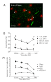
Neutrophils in microvascular injury after SAH. A: Representative image showing neutrophils in a brain section from an animal sacrificed 10 min after SAH. Note that some neutrophils (red) are within the collagen IV stained vessels (green; arrow heads) and others are present in the brain parenchyma (arrows). B: Area fractions of collagen IV and RECA-1 immunostaining in SAH and sham animals. Note that the area fraction of collagen IV and RECA-1 staining in SAH animals remained lower than the sham operated animals at all times examined. C: Numbers of neutrophils which are colocalized with collagen IV and RECA-1 after SAH. Filled circles: all neutrophils; filled squares: neutrophils that colocalized with collagen IV only; filled triangles: neutrophils that colocalized RECA-1 only. Open circles show all neutrophils in sham operated animals. Note that a greater number of neutrophils colocalized with collagen IV than with RECA-1 during the first 3 hours after SAH. Data are mean ± sem, N = 5 animals per time point and represent totals per whole coronal brain section * significantly different from sham operated animals (B) or from RECA-1 (C) at p < 0.05.
Figure 3
Figure 3
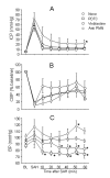
Early physiological changes after SAH: Animals were either untreated or were treated with vinblastine, anti PMN serum, or PDTC. ICP, CBF and BP were measured in real time from 20 minutes prior and 60 minutes post SAH (see methods). In A: note that ICP peak is similar in all groups but ICP decline in anti PMN group is significantly higher (25 ± 8 mmHg) than the untreated SAH animals (13 ± 1 mmHg). In B note that CAF fall and 60 minutes recovery is similar among animal groups. In C note that baseline BF and the transient increase in BP at SAH was similar among groups. There after BP decreased to lower levels in vinblastine and PDTC treated but not in anti PMN treated animals. Data are mean ± sem, N is 5-7 animals per treatment group. * significantly different at p < 0.05 from time matched untreated SAH animals.
Figure 4
Figure 4
Subarachnoid blood volume. Animals were either untreated or were treated with vinblastine, anti PMN serum, or PDTC and sacrificed one hour after SAH induction. The volume of blood surrounding circle of Willis was measured (see methods). Subarachnoid hemorrhage blood volume in anti PMN but not vinblastine and PDTC treated animals was significantly greater than the untreated SAH animals. Data are mean ± sem, N is 5 animals per treatment group. * significantly different at p < 0.05 from time matched untreated SAH animals.
Figure 5
Figure 5
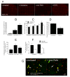
Pharmacological reduction of Neutrophils and their activity. A: Neutrophil staining in representative brain sections from untreated or vinblastine, anti PMN or PDTC treated animals sacrificed 1 hour after SAH. Note that fewer neutrophils are present in vinblastine and anti PMN treated animals and a large number are present in PDTC treated brains. B: Number of neutrophils in whole coronal brain sections. Values are % of untreated SAH animals. Neutrophils are decreased by vinblastin and anti-PMN treated and increased by PDTC treatment. C: RECA-1 (filled bars) and collagen IV (open bars) immunostaining following SAH. Values are area fractions in SAH animals as % of area fractions in sham-operated animals; both paramaters show trend or significant improvements in treated animals. D: Effect of PDTC treatment on the number of extravasated (parenchymal) neutrophils in SAH animals. Neutrophil extravasation is reduced by PDTC. E: Number of collagenase-positive profiles in treated SAH animals, given as % of values in untreated SAH animals. All three treatments reduce the extent of vascular collagenase activity. F: Effect of PDTC treatment on post-SAH intravascular tracer leakage. Values are area fractions of intravascular (closed bars) and extravascular (open bars) FITC-albumin deposits. G: Representative images of striatum showing vascular collagenase activity in untreated and anti PMN treated animals sacrificed at 1 hour after SAH. Arrows: collagen IV stained vessels (red) positive for collagenase activity (green). Data are mean ± sem. N = 5 animals per treatment group. * Significantly different at p < 0.05 from untreated SAH animals.

Similar articles

Cited by

References

    1. Yatsushige H, Ostrowski RP, Tsubokawa T, Colohan A, Zhang JH. Role of c-Jun N-terminal kinase in early brain injury after subarachnoid hemorrhage. J Neurosci Res. 2007;85:1436–1448. doi: 10.1002/jnr.21281. - DOI - PubMed
    1. Scholler K, Trinkl A, Klopotowski M, Thal SC, Plesnila N, Trabold R, Hamann GF, Schmid-Elsaesser R, Zausinger S. Characterization of microvascular basal lamina damage and blood-brain barrier dysfunction following subarachnoid hemorrhage in rats. Brain Res. 2007;1142:237–246. - PubMed
    1. Sehba FA, Mostafa G, Knopman J, Friedrich V Jr, Bederson JB. Acute alterations in microvascular basal lamina after subarachnoid hemorrhage. J Neurosurg. 2004;101:633–640. doi: 10.3171/jns.2004.101.4.0633. - DOI - PubMed
    1. Park KW, Metais C, Dai HB, Comunale ME, Sellke FW. Microvascular endothelial dysfunction and its mechanism in a rat model of subarachnoid hemorrhage. Anesth Analg. 2001;92:990–996. pp. 990-996.; - PubMed
    1. Friedrich V, Flores R, Muller A, Sehba FA. Escape of intraluminal platelets into brain parenchyma after subarachnoid hemorrhage. Neuroscience. 2010;165:968–975. doi: 10.1016/j.neuroscience.2009.10.038. - DOI - PMC - PubMed

Publication types

MeSH terms

LinkOut - more resources