Comparative Study
. 2011 Aug 19:4:162.
doi: 10.1186/1756-3305-4-162.

Secretory glands in cercaria of the neuropathogenic schistosome Trichobilharzia regenti - ultrastructural characterization, 3-D modelling, volume and pH estimations

Affiliations
Comparative Study

Secretory glands in cercaria of the neuropathogenic schistosome Trichobilharzia regenti - ultrastructural characterization, 3-D modelling, volume and pH estimations

Anna Ligasová et al. Parasit Vectors. .

Abstract

Background: Cercariae of schistosomes employ bioactive molecules for penetration into their hosts. These are released from specialized unicellular glands upon stimuli from host skin. The glands were previously well-described in the human pathogen Schistosoma mansoni. As bird schistosomes can also penetrate human skin and cause cercarial dermatitis, our aim was to characterize the architecture and ultrastructure of glands in the neurotropic bird schistosome Trichobilharzia regenti and compare it with S. mansoni. In the context of different histolytic enzymes used by these two species, we focused also on the estimations of gland volumes and pH in T. regenti.

Results: The architecture and 3-D models of two types of acetabular penetration glands, their ducts and of the head gland are shown here. We characterized secretory vesicles in all three gland types by means of TEM and confirmed accuracy of the models obtained by confocal microscopy. The results of two independent approaches showed that the glands occupy ca. one third of cercarial body volume (postacetabular glands ca. 15%, circumacetabular 12% and head gland 6%). The inner environment within the two types of acetabular glands differed significantly as evidenced by dissimilar ability to bind fluorescent markers and by pH value which was higher in circumacetabular (7.44) than in postacetabular (7.08) glands.

Conclusions: As far as we know, this is the first presentation of a 3-D model of cercarial glands and the first exact estimation of the volumes of the three gland types in schistosomes. Our comparisons between T. regenti and S. mansoni implied that the architecture and ultrastructure of the glands is most likely conserved within the family. Only minor variations were found between the two species. It seems that the differences in molecular composition have no effect on general appearance of the secretory cells in TEM. Fluorescent markers employed in this study, distinguishing between secretory vesicles and gland types, can be useful in further studies of mechanisms used by cercariae for host invasion. Results of the first attempts to estimate pH within schistosome glands may help further understanding of regulation of enzymatic activities present within the glands.


Figures

Figure 1
Figure 1
Glands of the cercaria of T. regenti. A, schematic figure of the cercaria with highlighted glands; postacetabular glands in green, circumacetabular glands in pink, head gland in blue. B, z-section from CM of cercarial head organ stained with Alexa Fluor® 488 and Cy3-azide; secretory vesicles of postacetabular ducts express bright fluorescence; head gland is in greenish grey. C, three-dimensional model of acetabular glands; Cy3-azide and Alexa Fluor® 488 stained cercaria was employed for the reconstruction; postacetabular glands are in green, circumacetabular glands in red; wide arrow shows acetabulum, thin arrow points to the strangulation of duct bundles entering muscular conus of the head organ (also see Additional File 1). D, visualization of cercarial circumacetabular glands by alizarin and fluorescence microscopy; cercaria was anaesthetized by Procain, acetabulum is exserted; marked are the posterior and anterior circumacetabular gland cells on the right side of cercaria. E, combination of autofluorescence and staining by FITC-phalloidin of the cercaria in CM; arrow points to the area where gland ducts enter the muscular conus; projection series. F, three-dimensional model of cercarial head gland; lobated cercarial head gland (in dark blue) with the three bundles of acetabular gland ducts on each side running through the head gland cell within the head organ (each bundle coloured separately). HG, head gland; dPA, ducts of postacetabular glands; CA-p and CA-a, posterior and anterior circumacetabular cells, respectively; PA and CA, postacetabular and circumacetabular glands, respectively; MC, muscle conus; A, acetabulum.
Figure 2
Figure 2
Formation of duct bundles anteriorly from the acetabulum in T. regenti cercaria. Longitudinal section in TEM; ducts of postacetabular and circumacetabular glands are grouped in two lateral bundles which are in a close contact at this point. CA, anterior pair of circumacetabular cells; dPA, postacetabular ducts; dCA, circumacetabular ducts; MC, posterior end of the muscular conus of the head organ; scale bar = 5 μm.
Figure 3
Figure 3
Electron micrographs of T. regenti gland ducts and openings. A, TEM of postacetabular and circumacetabular ducts reinforced by microtubules (arrows) and surrounded by common muscular layer; scale bar = 500 nm. B, TEM of the section through the apical part of cercarial head organ; yellow arrows show postacetabular ducts, red arrows show circumacetabular ducts and white arrows point to putative ducts of the head gland; note a sensory papilla adjoining with the gland duct opening covered by tegumental folds; scale bar = 5 μm; (also see Additional File 2). C, detail of four gland openings on the head organ of cercaria viewed by SEM; arrows point to sensory papillae in the proximity of the openings; scale bar = 5 μm. D, detail of a postacetabular gland duct in TEM leading to surface opening covered by tegumental folds (wide arrow) on the apex of cercaria, sensory papilla in the proximity of gland duct opening is pointed by thin arrow; scale bar = 2 μm. M, muscular layer; dCA and dPA, ducts of circumacetabular and postacetabular glands, respectively; SP, sensory papilla; HG, head gland; N, nucleus of the head gland.
Figure 4
Figure 4
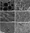
Details from transmission electron microscopy of secretory vesicles from particular glands of T. regenti cercaria. A-C, "classical" method of sample treatment; D-F, high-pressure freezing/freeze substitution method. A+D, postacetabular vesicles. B+E, circumacetabular vesicles. C+F, vesicles in the head gland. N, nuclei of muscle cells surrounded by the head gland cell. Scale bars = 500 nm.

Similar articles

Cited by

References

    1. Stirewalt MA, Kruidenier FJ. Activity of the acetabular secretory apparatus of cercariae of Schistosoma mansoni under experimental conditions. Exp Parasitol. 1961;11:191–211. doi: 10.1016/0014-4894(61)90026-1. - DOI - PubMed
    1. He Y, Yu Q, Yu P, Mao C, Hu Y. Penetration of Schistosoma japonicum cercaria into host skin. Chin Med J. 1990;103:34–44. - PubMed
    1. Neuhaus W. Biologie und Entwicklung von Trichobilharzia szidati n. sp. (Trematoda, Schistosomatidae), einem Erreger von Dermatitis bei Menschen. [Biology and development of Trichobilharzia szidati n. sp. (Trematoda, Schistosmatidae), a parasite causing dermatitis in man] Z Parasitenkd. 1952;15:203–266. - PubMed
    1. Müller V, Kimmig P. Trichobilharzia franki n. sp. - die Ursache fur Badedermatitiden in südwestdeutschen Baggerseen. [Trichobilharzia franki n. sp. - the cause of swimmer's dermatitis in southwest German dredged lakes] Appl Parasitol. 1994;35:12–31. - PubMed
    1. Horak P, Kolarova L, Dvorak J. Trichobilharzia regenti n. sp. (Schistosomatidae, Bilharziellinae), a new nasal schistosome from Europe. Parasite. 1998;5:349–357. - PubMed

Publication types