

Review
doi: 10.1186/1748-717X-6-97.
Guidelines for delineation of lymphatic clinical target volumes for high conformal radiotherapy: head and neck region
Affiliations
- PMID: 21854585
- PMCID: PMC3178490
- DOI: 10.1186/1748-717X-6-97
Item in Clipboard
Review
Guidelines for delineation of lymphatic clinical target volumes for high conformal radiotherapy: head and neck region
Radiat Oncol.
.
Abstract
The success of radiotherapy depends on the accurate delineation of the clinical target volume. The delineation of the lymph node regions has most impact, especially for tumors in the head and neck region. The purpose of this article was the development an atlas for the delineation of the clinical target volume for patients, who should receive radiotherapy for a tumor of the head and neck region. Literature was reviewed for localisations of the adjacent lymph node regions and their lymph drain in dependence of the tumor entity. On this basis the lymph node regions were contoured on transversal CT slices. The probability for involvement was reviewed and a recommendation for the delineation of the CTV was generated.
Figures
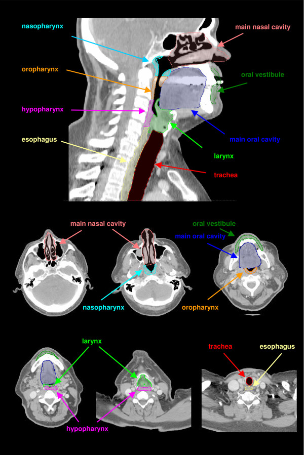
Anatomic head and neck regions contoured on a sagittal DRR and transversal CT slices.
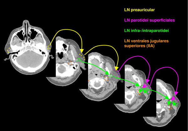
Lymph regions and drain contoured in transversal CT slices: LN parotidei superficiales (pink) and LN parotidei profundi subdivided into LN preauriculares (yellow) and LN infra-/intraparotidei (light green) [1.8 cm slice thickness].
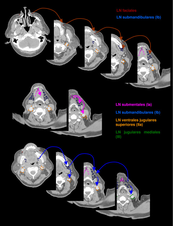
Lymph regions and drain contoured in transversal CT slices: LN buccales (brown), LN submentales (pink) and LN submandibulares (dark blue) [1.8 cm slice thickness].
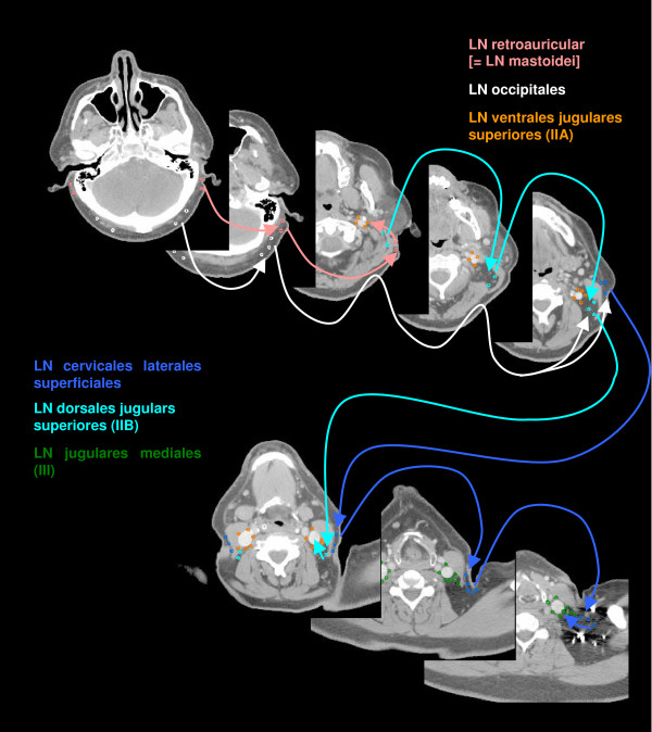
Lymph regions and drain contoured in transversal CT slices: LN occipitales (white), LN retroauriculares [ = LN mastoidei] (pink), LN cervicales laterales superficiales (medium blue) and LN dorsales jugulares superiores (cyan) [1.8 cm slice thickness].
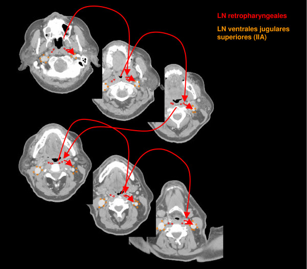
Lymph regions and drain contoured in transversal CT slices: LN retropharyngeales (red) [1 cm slice thickness].
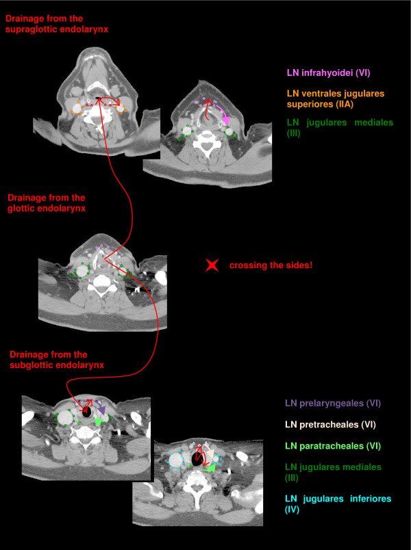
Lymph drainage from the endolarynx contoured in transversal CT slices (red arrows) to the LN infrahyoidei (pink), LN prelaryngeales (violet), LN pretracheales (light pink) and LN paratracheales (light green).
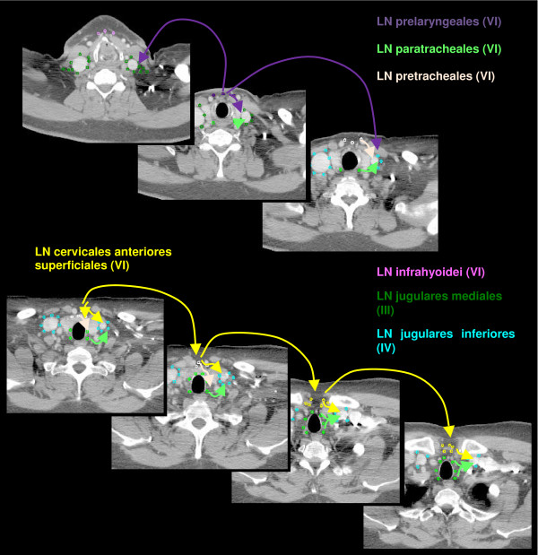
Lymph regions and drain contoured in transversal CT slices: LN cervicales anteriores superficiales (yellow) and LN cervicales anteriores profundi subdivided into LN infrahyoidei (pink), LN prelaryngeales (violet), LN pretracheales (light pink) and LN paratracheales (light green) [1 cm slice thickness].
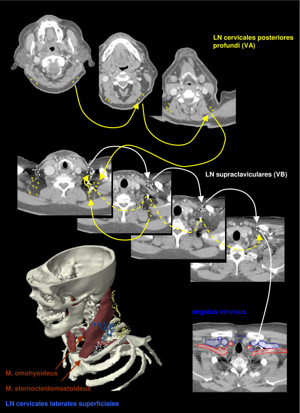
Lymph regions and drain contoured in transversal CT slices: LN cervicales posteriores profundi (yellow) and LN supraclaviculares (white).
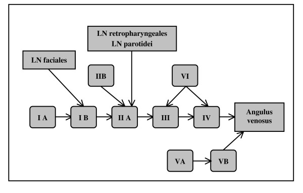
Schematic scheme of main direction of lymph node flow in the head and neck region.
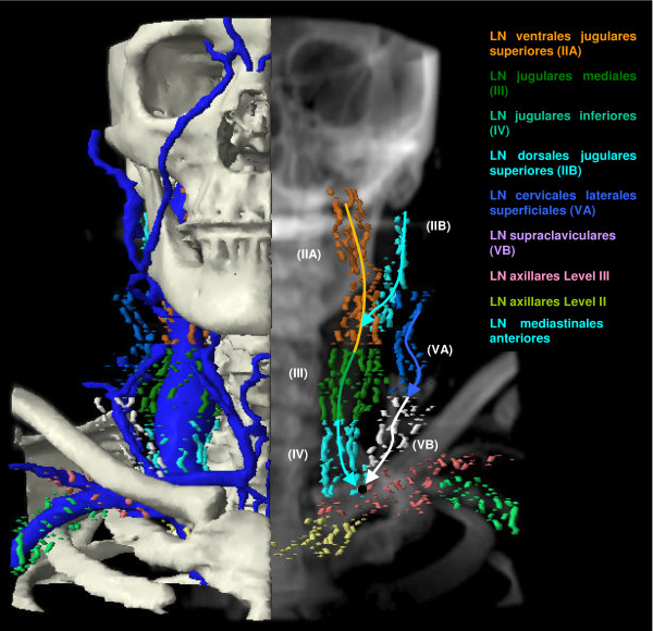
Coronar DRR with different lymph node regions, bones and veins. The black circle symbolises the angulus venosus.
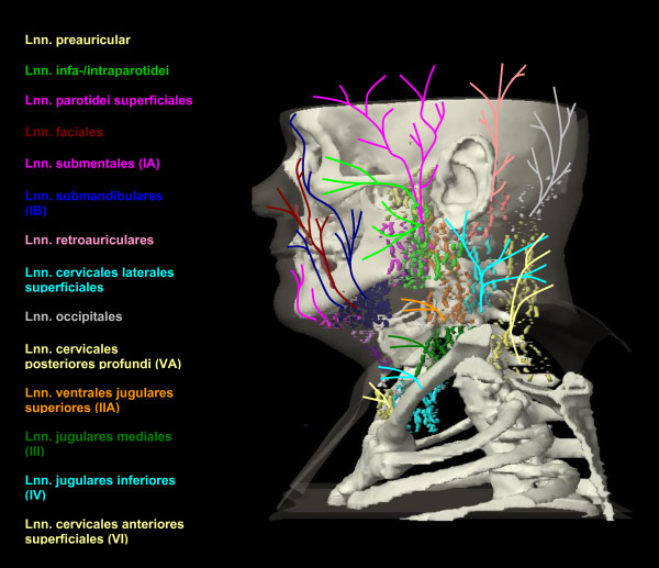
Lymph drain from the skin outlines as a schema on a capital view (for the systematic listing see Table 2): LN preauriculares (yellow, cranial), LN infra-/intraparotidei (light green), LN parotidei superficiales (pink, cranial), LN facials (brown), LN submentales (pink, ventral), LN submandibulares (dark blue), LN retroauriculares (rose), LN cervicales laterales superficiales (cyan cranio-dorsal), LN occipitals (grey), LN cervicales posteriores profundi (yellow, dorsal), LN ventrales jugulares superiores (orange), LN jugulares mediales (dark green), LN jugulares inferiores (cyan, caudo-ventral), LN cervicales anteriores superficiales (yellow, ventral).
References
-
- Keberle M, Ströbel P, Marx A, Hahn D, Hoppe F. CT determination of lymphocytic infiltration around head and neck squamous cell carcinomas may be a predictor of lymph node metastases. Eur Arch Otorhinolaryngol. 2003;260:558–564. - PubMed
Publication types
MeSH terms
LinkOut - more resources
Full Text Sources
Medical
