. 2011 Aug 19:9:117.
doi: 10.1186/1477-7827-9-117.

Developmental expression of p97/VCP (Valosin-containing protein) and Jab1/CSN5 in the rat testis and epididymis

Affiliations

Developmental expression of p97/VCP (Valosin-containing protein) and Jab1/CSN5 in the rat testis and epididymis

Sevil Cayli et al. Reprod Biol Endocrinol. .

Abstract

Background: The ubiquitin proteasome system (UPS) is a key player in regulating many cellular processes via proteasomal degradation of ubiquitinated proteins. Recently published data show that Jab1/CSN5 interacts with p97/VCP and controls the ubiquitination status of proteins bound to p97/VCP in mouse and human cells. However, coexpression of p97/VCP and Jab1/CSN5 in the developing rat testis and epididymis has not previously been studied.

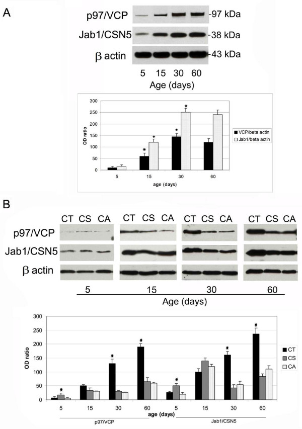
Methods: Testicular and epididymal tissues from 5-, 15-, 30-, and 60-day-old rats were examined by immunohistochemistry and Western blotting. Colocalisation of proteins was determined by immunofluorescence microscopy.

Results: In the 5-day-old rat testis, p97/VCP and Jab1/CSN5 were specifically expressed in gonocytes. The expression of p97/VCP and Jab1/CSN5 significantly increased at day 15 and was found in spermatogonia, Sertoli cells and spermatocytes. In 30- and 60-day-old rat testes, p97/VCP indicated moderate to strong expression in Sertoli cells, spermatogonia, round and elongating spermatids. However, moderate to weak expression was observed in spermatocytes. Jab1/CSN5 showed strong expression in spermatogonia and spermatocytes, while relatively moderate expression was observed in round and elongating spermatids in 30- and 60-day-old rat testes. In contrast, in the epididymis, the expression of both proteins gradually increased from 5 to 60 days of age. After rats reached 2 weeks of age, the expression of both proteins was mostly restricted to the basal and principal cells of the caput epididymis.

Conclusions: Our study suggests that p97/VCP and Jab1/CSN5 could be an important part of the UPS in the developing rat testis and epididymis and that both proteins may be involved in the regulation of spermatogenesis and epididymal epithelial functions.


Figures

Figure 1
Figure 1
Localisation of p97/VCP and Jab1/CSN5 in the developing rat testis. Immunohistochemistry was used to show the cellular localisation of p97/VCP and Jab1/CSN5 in the testis at days 5 (A, G), 15 (B, H), 30 (C, D, I), and 60 (E, F, J, K, L) after birth. The negative controls (A, C, G, H, L, inserts). A: Positive staining of p97/VCP expression was demonstrated in the nuclear and cytoplasmic regions of gonocytes (GC) at day 5. Interstitial cells (I) show weak immunoreactivity, and peritubuler cells (P) present no immunoreactivity. B: p97/VCP is highly expressed in spermatogonia (Sg), spermatocytes (Sc) and Sertoli cells (Se) at day 15. Some I show moderate immunostaining for p97/VCP. C, D: Elongating (eSt) and round (rSt) spermatids, Se and Sg show moderate to strong immunostaining for p97/VCP, while cytoplasm of Sc are moderately labelled with p97/VCP. E, F: In the adult testis, p97/VCP is weakly to moderately expressed in the cytoplasm of Sc, whereas Se, Sg, rSt and eSt are moderate to strong immunopositive for p97/VCP. G: Moderate Jab1/CSN5 expression is seen at day 5 in GC. H: Jab1/CSN5 is strongly expressed in Sg and Sc while Se exhibit moderate expression at day 15. I: Sg, Sc, eSt and Se show moderate to strong immunostaining for Jab1/CSN5. J-L: In the adult testis, Jab1/CSN5 is strongly expressed in Sg and Sc, while rSt and eSt show weak to moderate immunostaining for Jab1/CSN5. Roman numerals indicate stages of the seminiferous epithelial cycle. M: H-SCORE of the p97/VCP and Jab1/CSN5 immunostaining intensities in the developing rat testis. The data are represented as the means ± SEM. a: p < 0.05, day 5 vs. days 15, 30 and 60, b: day 15 vs. day 5 and day 30, c: day 30 vs. days 5 and 15. Scale bars: 50 μm.
Figure 2
Figure 2
Immunohistochemical analysis of Sertoli cells at day 5 (A), 15 (B), 60 (C) and colocalisation of p97/VCP - p27kip1 (D-F) and Jab1/CSN5 - p27kip1 (G-I) in rat testis. Cytokeratin 18 (CK) are detected in immature Sertoli cells (Se) at day 15 (A) and p27kip1 is observed in mature Sertoli cells (Se) of day 30 and 60 (B, C) in rat testis. Mature Sertoli cells (Se) are immunopositive for p27kip1 (green, D, G), p97/VCP (red, E) and Jab1/CSN5 (red, H), resulting in a yellow color (overlay, F, I). Scale bars: 25 μm.
Figure 3
Figure 3
Distribution of p97/VCP in different regions of the rat epididymis during postnatal development. p97/VCP immunopositivity is detected in the CT, CS and CA of the epididymis at days 5 (A, B, C), 15 (D, E, F), 30 (G, H, I) and 60 (J, K, L). p97/VCP expression is seen in the epithelial cells (EC) of the CT, CS and CA epididymis at day 5 (A, B, C). No significant staining is observed in the negative controls (at the corner of panels A-L). p97/VCP is localised both in the basal (BS) and principal (PC) cells of the epididymis at days 15, 30 and 60 (D-L). Of note, p97/VCP is also expressed in the apical layer of the cytoplasm in all epididymal regions at days 30 and 60 (G-L). Immunopositivity for p97/VCP is detected both in the principal cells and basal epithelial cells, while clear cells (C) show no immunoreactivity at day 60 (J-L). Spermatozoa (Sp) are weakly immunopositive for p97/VCP. CT: caput, CS: corpus, CA: cauda. Scale bars: 25 μm (A-C); scale bars: 50 μm (D-L).
Figure 4
Figure 4
Distribution of Jab1/CSN5 in different regions of the rat epididymis during postnatal development. Jab1/CSN5 immunopositivity is detected in the CT, CS and CA at days 5 (A, B, C), 15 (D, E, F), 30 (G, H, I) and 60 (J, K, L). Jab1/CSN5 is found in the cytoplasm of epithelial cells (EC) of the epididymis at day 5 (A, B, C). Both the basal (BS) and principal (PC) cells of the CT, CS and CA epididymis are found to be immunopositive for Jab1/CSN5 at days 15, 30 and 60 (D-L). In addition to the nuclear region of epithelial cells (EC), Jab1/CSN5 is highly expressed in the cytoplasm in the CT and CS epididymis at days 30 (G-I) and 60 (J-L). Spermatozoa (Sp) are weakly immunopositive for Jab1/CSN5 at day 60 (J-L). No significant staining is observed in the negative controls (at the upper corner of panels A-L). CT: caput, CS: corpus, CA: cauda. Scale bars: 25 μm (A-C); scale bars: 50 μm (D-L).
Figure 5
Figure 5
Double immunohistochemistry representing the colocalisation of p97/VCP and Jab1/CSN5 in the developing rat testis (A-D). Detection of Jab1/CSN5 (green), p97/VCP (red), and double-stained (yellow) positive cells merged with DAPI nuclear staining on day 5 (A), day 15 (B), day 30 (C) and day 60 (D). A: Day 5 gonocytes (GC) are double labelled with p97/VCP and Jab1/CSN5. B: Double-labelled spermatocytes (Sc), spermatogonia (Sg) and Sertoli cells (Se) are shown on day 15, and the nuclear regions of some spermatocytes are only positive for Jab1/CSN5. C: Jab1/CSN5 and p97/VCP positivity (yellow) is observed in spermatocytes (Sc), spermatogonia (Sg), Sertoli cells (Se) and elongating spermatids (eSt) on day 30. D: Double staining (yellow) is detected in the cytoplasm of spermatocytes (Sc), the nuclear region of spermatogonia (Sg) and elongating spermatids (eSt) and Sertoli cells (Se) in the 60-day-old rat testis. Scale bars: 25 μm.
Figure 6
Figure 6
Colocalisations of Jab1/CSN5 and p97/VCP in the developing rat epididymis (A-D). Detection of Jab1/CSN5 (green), p97/VCP (red), and double (yellow) positive cells merged with DAPI nuclear staining on day 5 (A), 15 (B), 30 (C) and 60 (D) CS epididymis. Both the basal (BS) and principal cells (PC) of CS epididymis are immunopositive for Jab1/CSN5 (green) and for p97/VCP (red), resulting a yellow color. Spermatozoa (Sp) are weakly immunopositive for Jab1/CSN5 and p97/VCP on day 60. Scale bars: 25 μm (A), 50 μm (B-D).
Figure 7
Figure 7
Western blot analysis of p97/VCP and Jab1/CSN5 in the developing rat testis (A) and epididymis (B). A: p97/VCP (97 kDa) and Jab1/CSN5 (38 kDa) were detected by western blotting. β-actin (43 kDa) was used as loading control. Immunoblot bands were quantified by an optical densitometer. The OD (optical density) values of the p97/VCP and Jab1/CSN5 bands were normalised to the OD values of β-actin bands. The data in the graphs are presented as the means ± SEM. The expression of p97/VCP and Jab1/CSN5 is significantly increased from days 5 to 30 in the testis (Asterisks indicate, p < 0.05); however, no differences are observed between days 30 and 60. B: p97/VCP and Jab1/CSN5 expression levels are significantly higher in the CT epididymis than the CS and CA epididymis at days 30 and 60. Asterisks indicate p < 0.05, days 5 CS vs. days 5 CA and CT epididymis, days 30 and 60 CT vs. days 30 and 60 CA and CS epididymis. CT: caput, CS: corpus, CA: cauda.

Similar articles

Cited by

References

    1. de Kretser DM. Clinical male infertility. I. Prevalence of and progress in understanding male infertility. Reprod Fertil Dev. 1994;6:3–8. doi: 10.1071/RD9940003. - DOI - PubMed
    1. Kierszenbaum AL. Mammalian spermatogenesis in vivo and in vitro: a partnership of spermatogenic and somatic cell lineages. Endocr Rev. 1994;15:116–134. - PubMed
    1. Clermont Y, Perey B. Quantitative study of the cell population of the seminiferous tubules in immature rats. Am J Anat. 1957;100:241–267. doi: 10.1002/aja.1001000205. - DOI - PubMed
    1. Cooper TG. Interactions between epididymal secretions and spermatozoa. J Reprod Fertil Suppl. 1998;53:119–136. - PubMed
    1. Orgebin-Crist MC. Studies on the function of the epididymis. Biol Reprod. 1969;1(Suppl 1):155–175. - PubMed

Publication types

LinkOut - more resources