. 2011 Aug 19:12:421.
doi: 10.1186/1471-2164-12-421.

The tammar wallaby major histocompatibility complex shows evidence of past genomic instability

Affiliations

The tammar wallaby major histocompatibility complex shows evidence of past genomic instability

Hannah V Siddle et al. BMC Genomics. .

Abstract

Background: The major histocompatibility complex (MHC) is a group of genes with a variety of roles in the innate and adaptive immune responses. MHC genes form a genetically linked cluster in eutherian mammals, an organization that is thought to confer functional and evolutionary advantages to the immune system. The tammar wallaby (Macropus eugenii), an Australian marsupial, provides a unique model for understanding MHC gene evolution, as many of its antigen presenting genes are not linked to the MHC, but are scattered around the genome.

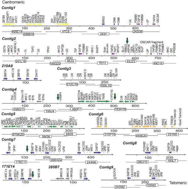
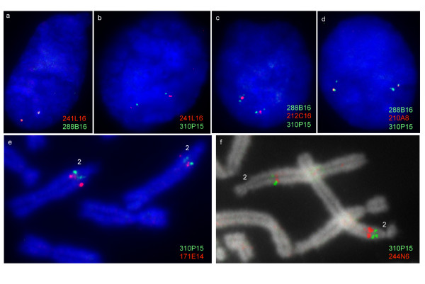
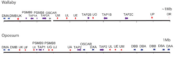
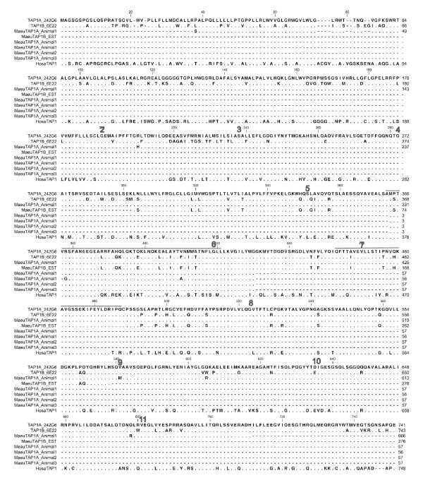
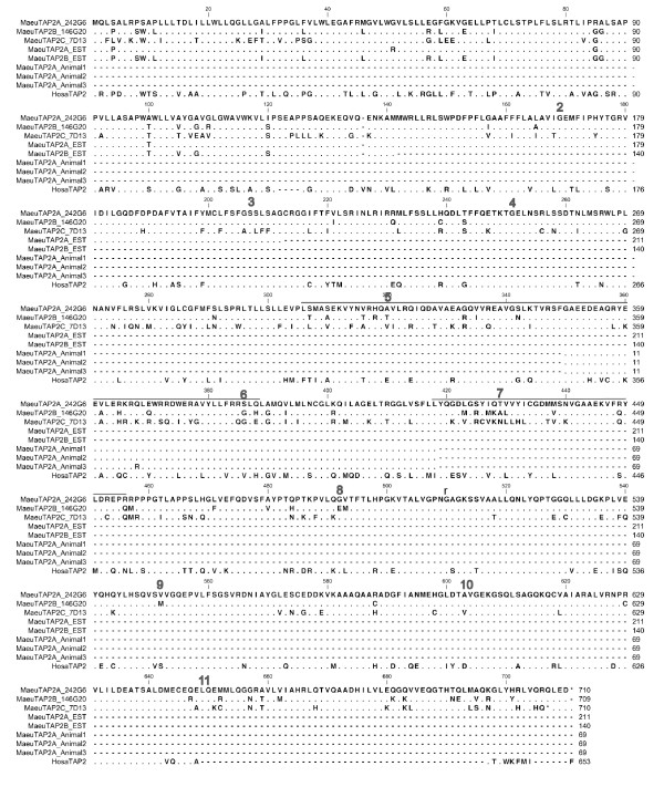
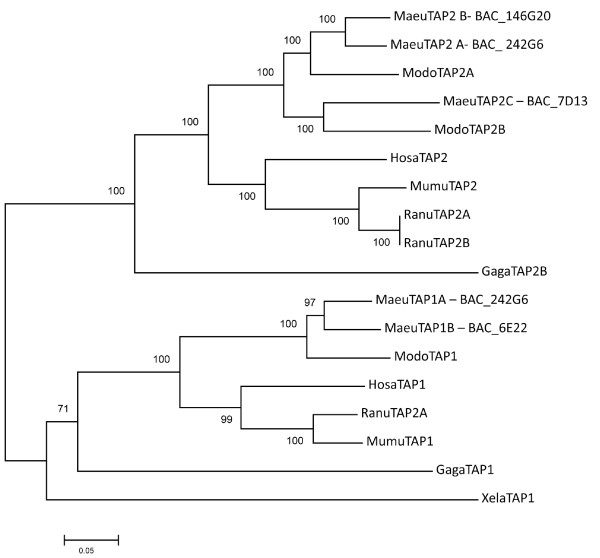
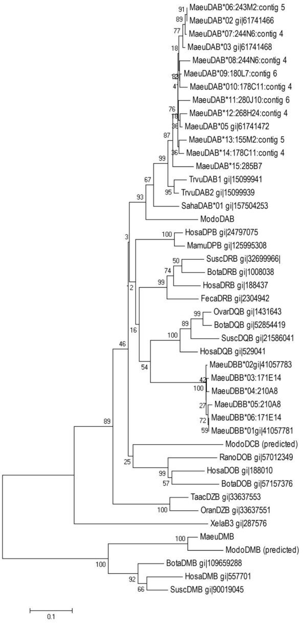
Results: Here we describe the 'core' tammar wallaby MHC region on chromosome 2q by ordering and sequencing 33 BAC clones, covering over 4.5 MB and containing 129 genes. When compared to the MHC region of the South American opossum, eutherian mammals and non-mammals, the wallaby MHC has a novel gene organization. The wallaby has undergone an expansion of MHC class II genes, which are separated into two clusters by the class III genes. The antigen processing genes have undergone duplication, resulting in two copies of TAP1 and three copies of TAP2. Notably, Kangaroo Endogenous Retroviral Elements are present within the region and may have contributed to the genomic instability.

Conclusions: The wallaby MHC has been extensively remodeled since the American and Australian marsupials last shared a common ancestor. The instability is characterized by the movement of antigen presenting genes away from the core MHC, most likely via the presence and activity of retroviral elements. We propose that the movement of class II genes away from the ancestral class II region has allowed this gene family to expand and diversify in the wallaby. The duplication of TAP genes in the wallaby MHC makes this species a unique model organism for studying the relationship between MHC gene organization and function.


Figures

Figure 1
Figure 1
Diagram of the organization of the wallaby MHC with gene annotation. Colour code for genes; yellow - extended class II, blue - class II, red - class I, purple - antigen processing genes, pink - olfactory receptors, grey - pseudogenes. The overlapping BACs are indicated by black lines below the annotation. BACs not assembled into a contig are indicated by the BAC name. The KERV fragments are indicated by thick black arrows. An OSCAR fragment is indicated with an arrow.
Figure 2
Figure 2
Metaphase and interphase FISH showing the location of anchored BACs in order to order the contigs. (a-d) FISH on tammar walaby interphase nuclei showing (a) co-localization of contig 2 (BAC_241L16 in red) and contig 1 (BAC_288B16 in green), b) the positions of contig 2 (BAC_241L16 in red) and contig 6 (BAC_310P15 in green) relative to one another, c) the position of BAC_212C16 relative to contig 1 (BAC_288B16) and contig 6 (BAC_310P15), BAC_212C16 is midway between the two contigs, d) the position of BAC_210A8 relative to contig 1 (BAC_288B16) and contig 6 (BAC_310P15), BAC_210A8 is closer to BAC_288B16 than BAC_310P15. e) Metaphase FISH showing that contig 4 (BAC_244N6 in red) is telomeric to contig 6 (BAC_310P15 in green), f) Metaphase FISH showing that BAC_171E14 in red is approximately 1 Mb telomeric to contig 6 (BAC_310P15 in green).
Figure 3
Figure 3
Schematic comparison of the opossum MHC class I/II region and the putative class I/II region in the tammar wallaby. Colour code for genes; blue-class II, red-class I, purple-antigen processing genes, grey-pseudogenes. Each 100 Kb is represented by a short black line.
Figure 4
Figure 4
Alignment of TAP1 genes from the wallaby and expressed transcripts isolated from spleen, blood and EST library. Amino acid alignment of TAP1 sequences. TAP1A_242G6 and TAP1B_6E22 are sourced from BACs in Contigs 1 and 2 respectively. MaeuTAP1A_Animal1 was sourced from a the spleen of animal1, TAP1B_EST is sourced from a mixed tissue EST library. MaeuTAP1A_Animal1, MaeuTAP1A_Animal2 and MaeuTAP1A_Animal3 were sourced from blood samples from Animals 1, 2 and 3 respectively. HosaTAP1 is included for comparison. Dashes indicate missing sequence, while dots indicate conserved residues. Exon boundaries are indicated by a number above the first residue of the exon. Residues thought to interact with the peptide are indicated by a line above the sequence.
Figure 5
Figure 5
Alignment of TAP2 genes from the wallaby and expressed transcripts isolated from spleen, blood and EST library. Amino acid alignment of TAP2 sequences. TAP2A_242G6, TAP2B_146G20 and TAP2C_6E22 were sourced from BACs in Contigs 1 and 2. MaeuTAP2A_EST and MaeuTAP2B_EST were sourced from a mixed tissue EST library. MaeuTAP2A from Animals 1, 2 and 3 were sourced from blood samples. HosaTAP2 is included for comparison. Dashes indicate missing sequence, while dots indicate conserved residues. Exon boundaries are indicated by a number above the first residue of the exon. Residues thought to interact with the peptide are indicated by a line above the sequence.
Figure 6
Figure 6
Neighbour-joining phylogenetic tree showing the relationship between TAP1 and TAP2 genes from the wallaby and other vertebrates. Analysis was performed on the full amino acid sequence for each gene.
Figure 7
Figure 7
Neighbour joining phylogenetic tree showing the relationship between mammalian class II B chain genes. Analysis was performed on the amino acid sequence from the β2 domain only.

References

    1. Shiina T, Inoko H, Kulski JK. An update of the HLA genomic region, locus information and disease associations: 2004. Tissue Antigens. 2004;64(6):631–649. doi: 10.1111/j.1399-0039.2004.00327.x. - DOI - PubMed
    1. Carrington M, Nelson GW, Martin MP, Kissner T, Vlahov D, Goedert JJ, Kaslow R, Buchbinder S, Hoots K, O'Brien SJ. HLA and HIV-1: heterozygote advantage and B*35-Cw*04 disadvantage. Science. 1999;283(5408):1748–1752. doi: 10.1126/science.283.5408.1748. - DOI - PubMed
    1. Madsen T, Ujvari B. MHC class I variation associates with parasite resistance and longevity in tropical pythons. J Evol Biol. 2006;19(6):1973–1978. doi: 10.1111/j.1420-9101.2006.01158.x. - DOI - PubMed
    1. Kulski JK, Shiina T, Anzai T, Kohara S, Inoko H. Comparative genomic analysis of the MHC: the evolution of class I duplication blocks, diversity and complexity from shark to man. Immunol Rev. 2002;190:95–122. doi: 10.1034/j.1600-065X.2002.19008.x. - DOI - PubMed
    1. Kelley J, Walter L, Trowsdale J. Comparative genomics of major histocompatibility complexes. Immunogenetics. 2005;56(10):683–695. doi: 10.1007/s00251-004-0717-7. - DOI - PubMed

Publication types

Associated data