. 2011 Aug 19;11(1):29.
doi: 10.1186/1475-2867-11-29.

The effect of the histone deacetylase inhibitor M344 on BRCA1 expression in breast and ovarian cancer cells

Affiliations

The effect of the histone deacetylase inhibitor M344 on BRCA1 expression in breast and ovarian cancer cells

Johanne I Weberpals et al. Cancer Cell Int. .

Abstract

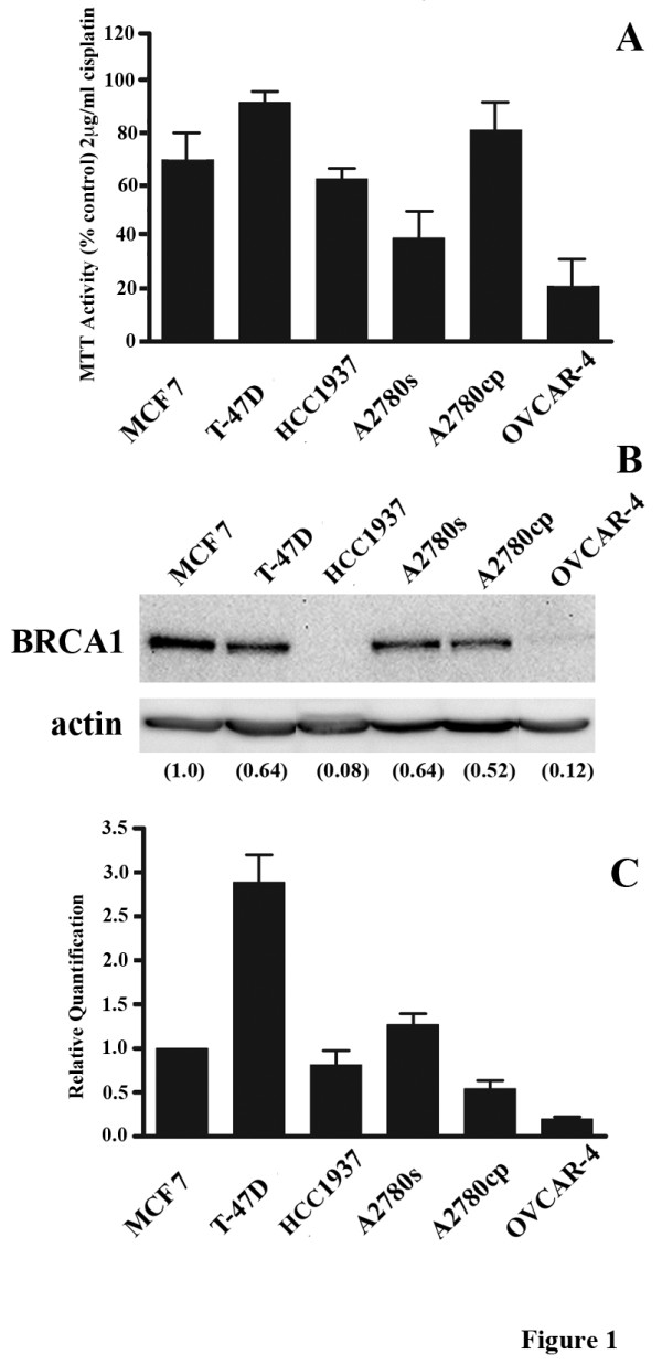
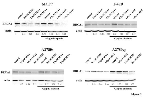
Background: The inhibition of Breast Cancer 1 (BRCA1) expression sensitizes breast and ovarian cancer cells to platinum chemotherapy. However, therapeutically relevant agents that target BRCA1 expression have not been identified. Our recent report suggested the potential of the histone deacetylase (HDAC) inhibitor, M344, to inhibit BRCA1 expression. In this study, we further evaluated the effect of M344 on BRCA1 mRNA and protein expression, as well as its effect on cisplatin-induced cytotoxicity in various breast (MCF7, T-47D and HCC1937) and ovarian (A2780s, A2780cp and OVCAR-4) cancer cell lines.

Results: With the addition of M344, the platinum-sensitive breast and ovarian cancer cell lines that displayed relatively high BRCA1 protein levels demonstrated significant potentiation of cisplatin cytotoxicity in association with a reduction of BRCA1 protein. The cisplatin-resistant cell lines, T-47D and A2780s, elicited increased cytotoxicity of cisplatin with M344 and down regulation of BRCA1 protein levels. A2780s cells subjected to combination platinum and M344 treatment, demonstrated increased DNA damage as assessed by the presence of phosphorylated H2A.X foci in comparison to either treatment alone. Using Chromatin Immunoprecipitation, A2780s and MCF7 cells exposed to M344 alone and in combination with cisplatin, did not demonstrate enhanced acetylated Histone 4 at the BRCA1 promoter, suggesting an indirect effect on this promoter.

Conclusions: The enhanced sensitivity of HDAC inhibition to platinum may be mediated through a BRCA1-dependent mechanism in breast and ovarian cancer cells. The findings of this study may be important in the future design of clinical trials involving HDAC inhibitors using BRCA1 as a tumour biomarker.


Figures

Figure 1
Figure 1
Cisplatin sensitivity and BRCA1 expression in the breast and ovarian cancer derived cell lines evaluated in this study. A) MTT cell viability assays comparing the responses of a panel of cell lines to 2 μg/ml cisplatin. Cell viability was assayed with the activity of untreated cells taken to be 100%. Values represent the mean +/- SEM of three separate experiments. B) Western blot analysis of basal expression levels of BRCA1 protein in a panel of cell lines. Actin was employed as a loading control. Numbers indicate protein densitometry readings with MCF7 used as the calibrator and set to 1.0. The experiment was repeated with similar results. C) Basal levels of BRCA1 mRNA analyzed by RT-PCR. Relative expression for each cell line was calculated following normalization to GAPDH levels and then further normalized to MCF-7 for ease of comparison and expressed as the mean +/- SEM of three separate experiments.
Figure 2
Figure 2
Effect of M344 and cisplatin treatments on the levels of BRCA1 mRNA in our panel of cell lines. BRCA1 mRNA levels were analyzed by RT-PCR following 24 hrs of treatment with 0.5, 1.0, or 5.0 μM M344 either alone or in combination with 2 μg/ml cisplatin. DMSO is the solvent control for the M344 treatment and 2 μg/ml cisplatin is the control for the combination. Values represent the mean +/- SEM of three separate experiments. Statistically significant differences, when present, between 5 μM M344 and DMSO control or 2 μg/ml cisplatin + 5 μM M344 and 2 μg/ml cisplatin alone are indicated by * where P < 0.05, ** where P < 0.01, and *** where P < 0.001.
Figure 3
Figure 3
Effect of M344 and cisplatin treatments on the levels of BRCA1 protein expression in our panel of cell lines. Western blot analysis of BRCA1 following 24 hrs of treatment with 0.5, 1.0, or 5.0 μM M344 either alone or in combination with 2 μg/ml cisplatin. DMSO is the solvent control for the M344 treatment and 2 μg/ml cisplatin is the control for the combination treatment. The experiment was repeated with similar results. Densitometry readings are normalized to the DMSO solvent control.
Figure 4
Figure 4
Analysis of the cytotoxic and apoptotic effects of the combination of M344 and cisplatin inn this cell line panel. A) MTT cell viability assays comparing the responses of a panel of cell lines to 1 μM M344 alone, 2 μg/ml cisplatin alone or both treatments in combination for 48 hrs. Cell viability was assayed with the activity of untreated cells taken to be 100%. Values represent the mean +/- SEM of three separate experiments. Differences between treatment with cisplatin alone versus treatment with the cisplatin and M344 combination were analyzed using paired T-test analyses. * indicates a significant difference where P < 0.001. B) Percentage of apoptotic cells in the A2780s cell line following 24 hrs of treatment and A2780cp cell line following 48 hrs treatment with 1.0 μM M344 alone or in combination with 2 μg/ml cisplatin as assessed by flow cytometry. The experiment was done in three replicates with similar results.
Figure 5
Figure 5
Evaluating the effect of M344, cisplatin and their combination on the formation of γH2A.X foci. A) Phase contrast images of A2780s cells following 24 hrs of treatment with 5.0 μM M344 alone or in combination with 2 μg/ml cisplatin. B) A2780s cells subjected to combination platinum and M344 24 hr treatment demonstrates increased levels γH2A.X foci detected by immunoflourescence. Repeat of the experiment showed similar findings.
Figure 6
Figure 6
ChIP analysis of the levels of acetylated histones at the BRCA1 promoter following M344 and cisplatin treatments. A) MCF7 and B) A2780s cells treated with 5.0 μM M344 in combination with 2 μg/ml cisplatin for 24 hrs show reduced amounts of BRCA1 promoter DNA bound to acetylated histone 4 (AcH4). Real-time PCR products were run on a 1.6% agarose gel. "Input" controls: untreated; "No Ab" controls: incubation with agarose beads in the absence of αAcH4; and "αAcH4": incubation with agarose beads and αAcH4. The experiment was repeated twice with similar results.

Similar articles

Cited by

References

    1. Jemal A, Siegel R, Xu J, Ward E. Cancer Statistics, 2010. CA Cancer J Clin. 2000;60:277–300. - PubMed
    1. Hall JM, Lee MK, Newman B, Morrow JE, Anderson LA, Huey B, King MC. Linkage of early-onset familial breast cancer to chromosome 17q21. Science. 1990;250:1684–9. doi: 10.1126/science.2270482. - DOI - PubMed
    1. Miki Y, Swensen J, Shattuck-Eidens D, Futreal PA, Harshman K, Tavtigian S, Liu Q, Cochran C, Bennett LM, Ding W. et al.A strong candidate for the breast and ovarian cancer susceptibility gene BRCA1. Science. 1994;266:66–71. doi: 10.1126/science.7545954. - DOI - PubMed
    1. Thrall M, Gallion HH, Kryscio R, Kapali M, Armstrong DK, DeLoia JA. BRCA1 expression in a large series of sporadic ovarian carcinomas: a Gynecologic Oncology Group study. Int J Gynecol Cancer. 2006;16(Suppl 1):166–71. - PubMed
    1. Turner N, Tutt A, Ashworth A. Hallmarks of 'BRCAness' in sporadic cancers. Nat Rev Cancer. 2004;4:814–9. doi: 10.1038/nrc1457. - DOI - PubMed

LinkOut - more resources