. 2011 Oct;179(4):1988-2000.
doi: 10.1016/j.ajpath.2011.06.044. Epub 2011 Aug 18.

Modeling oculopharyngeal muscular dystrophy in myotube cultures reveals reduced accumulation of soluble mutant PABPN1 protein

Affiliations

Modeling oculopharyngeal muscular dystrophy in myotube cultures reveals reduced accumulation of soluble mutant PABPN1 protein

Vered Raz et al. Am J Pathol. 2011 Oct.

Abstract

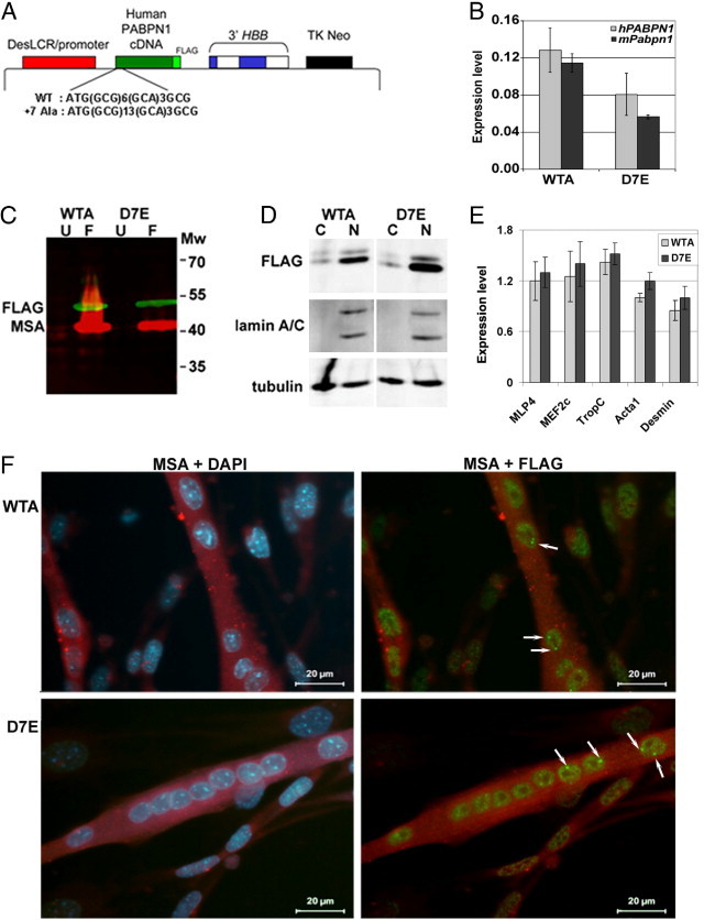
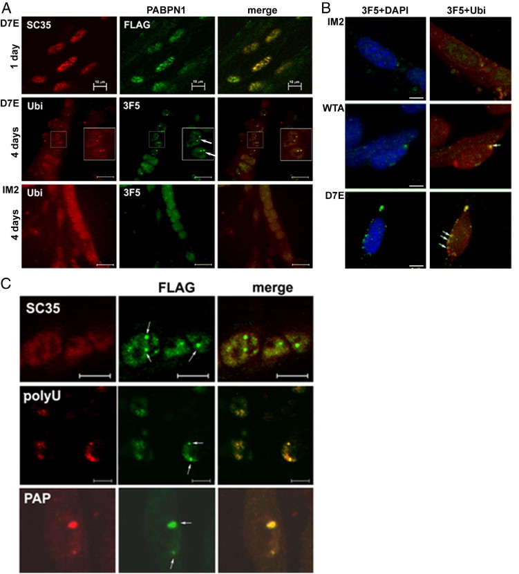
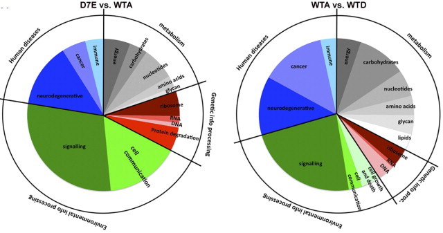
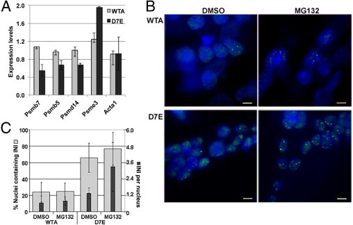
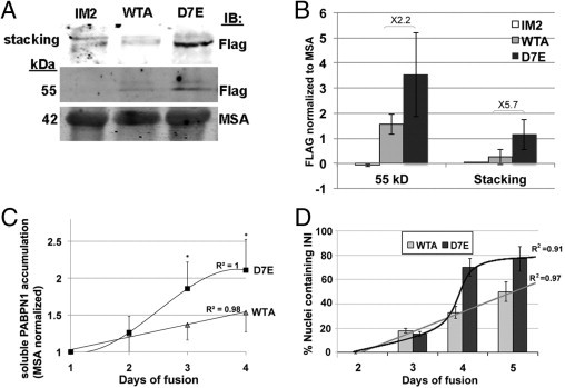
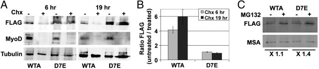
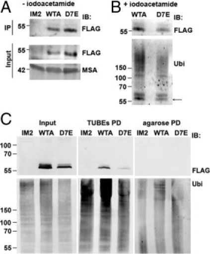
Oculopharyngeal muscular dystrophy (OPMD) is an autosomal dominant disease caused by an alanine tract expansion mutation in poly(A) binding protein nuclear 1 (expPABPN1). To model OPMD in a myogenic and physiological context, we generated mouse myoblast cell clones stably expressing either human wild type (WT) or expPABPN1 at low levels. Transgene expression is induced on myotube differentiation and results in formation of insoluble nuclear PABPN1 aggregates that are similar to those observed in patients with OPMD. Quantitative analysis of PABPN1 in myotube cultures revealed that expPABPN1 accumulation and aggregation is greater than that of the WT protein. We found that aggregation of expPABPN1 is more affected than WT PABPN1 by inhibition of proteasome activity. Consistent with this, in myotube cultures expressing expPABPN1, deregulation of the proteasome was identified as the most significantly perturbed pathway. Differences in the accumulation of soluble WT and expPABPN1 were consistent with differences in ubiquitination and rate of protein turnover. This study demonstrates, for the first time to our knowledge, that, in myotubes, the ratio of soluble/insoluble expPABPN1 is significantly lower compared with that of the WT protein. We suggest that this difference can contribute to muscle weakness in OPMD.


Figures

Figure 1
Figure 1
Stably transfected myoblast cell lines expressing either WT or mutant 7Ala-expanded PABPN1. A: Illustration of the muscle-specific DesLCR-EV expression vector containing cDNAs (green box) coding for either human WT (10 Ala; WTA) or 7Ala-expanded (17Ala; D7E) PABPN1. The PABPN1 in both cases is tagged at its C-terminus with the FLAG epitope (DYKDDDDK). The 5′ sequence of the PABPN1 cDNA showing the WT and the mutated poly-Ala track (GCG, GCA) is indicated. The DesLCR/promoter, a muscle-specific element, drives transgene expression in myotubes. The 3′ half of HBB at the 3′ of the expression cassette confers efficient processing, transport, and stability of the mRNA. A neomycin-resistance gene under the control of an HSV-TK promoter allows selection of stably transfected clones. B: A histogram showing expression levels of murine mPabpn1 and human hPABPN1 mRNA expression in WTA and D7E that were induced for myotube fusion for 4 days. The expression level is calculated from ΔΔCT values after normalization to DES and to IM2 parental cells. Error bars are calculated from three replicates. C: PABPN1-FLAG expression in myotubes. Western blot analysis of total protein extracts of unfused (U) and 4 days after myoblast fusion (F) of WTA and D7E cell lines. Molecular weights (Mw) are indicated in kDa. D: Western blot analysis of cytoplasmic (C) and nuclear (N) fractions isolated from 4-day fused WTA and D7E cell lines. Anti-lamin A marks the nuclear fraction, and tubulin was used as a loading control for both fractions. The blot is a representative of three independent experiments. E: RT-qPCR analysis of muscle differentiation markers in WTA and D7E cell lines. Expression levels were calculated from ΔΔCT values after normalization to murine Hrpt and to IM2 parental cells. Error bars are calculated from three replicates. F: IHC of PABPN1-FLAG in WTA and D7E cell lines 4 days after fusion. PABPN1-FLAG is detected with an anti-FLAG antibody, and differentiated myotubes are detected with an MSA antibody. Nuclei are counterstained with DAPI. Arrows show INIs.
Figure 2
Figure 2
INIs are formed in WTA and D7E cells lines. A: INIs are formed in D7E myotubes but not in myoblasts or in IM2 myotubes. In unfused cells (day 1), PABPN1-FLAG colocalizes with SC35, indicating its localization in speckles. Scale bar = 10 μm. In 4-day myotube cultures of D7E, but not of IM2 PABPN1, fluorescent foci, labeled with VHH-3F5, colocalize with ubiquitin (Ubi). Scale bar = 20 μm. Original magnification, ×1.5 (of nuclei with INIs, highlighted in boxes). B: INIs in D7E and WTA are KCl resistant. At 4 days after fusion, cultures were treated with 1 mol/L KCl before IHC with VHH-3F5 (green) and Ubi (red). Nuclei were counterstained with DAPI. Arrows, INI. Scale bar = 5 μm. C: INIs colocalize with polyU and poly(A) polymerase (PAP) but not with SC35. Myotubes of D7E were coimmunolabeled with SC35, polyU, or PAP. The PABPN1-FLAG protein is labeled with an anti-FLAG antibody. Scale bar = 5 μm. Arrows, INI.
Figure 3
Figure 3
Transcriptome study in D7E and WTA myotubes. Pie chart analysis of KEGG pathways that are deregulated in D7E (left) or WTA (right) myotubes. The inner pie shows the subcategories, and the outer ring shows the major categories.
Figure 4
Figure 4
Proteasome down-regulation is associated with protein aggregation in D7E. A: RT-qPCR analysis of proteasome-encoding genes in 4-day myotube cultures of WTA and D7E. Expression levels were calculated from ΔΔCT values after normalization to murine Hrpt and to IM2 parental cells. Error bars are calculated from two biological and six technical replicates. B: The effect of 5 μmol/L MG132 treatment on PABPN1 aggregation. Myotube cultures of WTA and D7E at 4 days after fusion were treated with 5 μmol/L MG132 for 8 hours. Dimethylsulfoxide (DMSO) treatment was used as a control. Protein aggregation and INI formation are detected by immunofluorescence using an anti-FLAG antibody, and nuclei are counterstained with DAPI. Scale bar = 5 μm. C: Histograms show the percentage of nuclei containing PABPN1 INI (gray bars) and the average number of INIs per nucleus (black bars). SDs represent 10 myotubes containing 90 nuclei in total.
Figure 5
Figure 5
expPABPN1 aggregation potency is higher than WT-PABPN1 in myotubes. A: Western blot analysis of total protein extracts isolated from IM2, WTA, and D7E myotubes. PABPN1-FLAG is detected with an anti-FLAG antibody. Aggregated PABPN1-FLAG is retained in the stacking gel, and the soluble protein migrates at 53 kDa. Muscle skeletal actin (MSA) is used as a loading control. B: Quantification of PABPN1-FLAG accumulation in IM2, WTA, and D7E myotubes at 4 days after fusion. The histogram shows the FLAG signal after normalizing to actin. Averages are calculated from six independent experiments. C: Quantification of soluble PABPN1-FLAG protein accumulation in WTA and D7E during cell fusion. Values show the FLAG signal normalized to MSA. Averages are calculated from six replicates. *P < 0.05. Linear or sigmoid curve fits of PABPN1-FLAG accumulation in WTA or D7E, respectively (R2 values are indicated). D: INI formation in WTA and D7E cell lines during myoblast cell fusion. INIs were visualized with an anti-FLAG antibody. Histograms show the percentage of nuclei containing PABPN1 INI from 80 to 100, counted in 20 myotubes at 2, 3, 4, and 5 days after fusion. A linear or sigmoid curve fits INI formation in WTA or D7E, respectively (R2 values are indicated).
Figure 6
Figure 6
Soluble expPABPN1 protein has slow turnover, and its accumulation is affected by proteasome inhibition. A: Cyclohexamide treatment (Chx; 10 μmol/L) of 3-day myotube cultures was conducted for 6 or 19 hours. Extracts of soluble protein were resolved by SDS-PAGE. The blot image shows the 53-kDa PABPN1-FLAG. MyoD is a control for cyclohexamide treatment, and tubulin is a loading control. B: Histograms show the ratio of FLAG signal between cyclohexamide untreated and treated WTA and D7E myotube cultures. SDs represent two experiments. C: Myotube cultures of WTA and D7E at 4 days after fusion were treated with 5 μmol/L MG132 for 8 hours. Dimethylsulfoxide treatment was used as a control. The soluble PABPN1 protein was detected by using Western blot analysis. PABPN1-FLAG was detected with anti-FLAG antibody; MSA is used as a fusion marker and as a loading control. The fold change of the FLAG signal between treated and untreated WTA and D7E myotubes is indicated.
Figure 7
Figure 7
Ubiquitination of soluble PABPN1. Ubiquitination of soluble PABPN1 protein was compared between extracts of soluble proteins in the absence (A) or presence (B) of iodoacetamide. Protein extracts were prepared from myotube cultures of WTA, D7E, or IM2 at 4 days after fusion. Aliquots were used for immunoprecipitation (IP) with rabbit anti-FLAG antibody. Western blot analysis was conducted with a mouse anti-FLAG antibody. Protein input is indicated in the input fraction, and MSA is used as a loading control. FLAG-immunoprecipitated ubiquitinated proteins were detected with anti-ubiquitin antibody (Ubi) (B). Arrow, PABPN1. C: Detection of ubiquitinated PABPN1 in D7E and WTA myotubes was performed by TUBE pull down (PD). Myotubes were mock treated (-MG132) or treated with 5 μmol/L MG132 for 8 hours (+MG132). Protein aliquots are shown in the input fraction. Pull down of naked agarose beads and extracts from myotubes of IM2 parental cells were used as negative controls. Immunoblot (IB) of PABPN1 was performed with mouse anti-FLAG antibody, and ubiquitinated proteins were detected with an anti-uniquitin (UBi) antibody.

Similar articles

Cited by

References

    1. Cummings C.J., Zoghbi H.Y. Fourteen and counting: unraveling trinucleotide repeat diseases. Hum Mol Genet. 2000;9:909–916. - PubMed
    1. Albrecht A., Mundlos S. The other trinucleotide repeat: polyalanine expansion disorders. Curr Opin Genet Dev. 2005;15:285–293. - PubMed
    1. Brown L.Y., Brown S.A. Alanine tracts: the expanding story of human illness and trinucleotide repeats. Trends Genet. 2004;20:51–58. - PubMed
    1. Ross C.A., Poirier M.A. Protein aggregation and neurodegenerative disease. Nat Med. 2004;10(Suppl):S10–S17. - PubMed
    1. Brais B., Bouchard J.P., Xie Y.G., Rochefort D.L., Chretien N., Tome F.M., Lafreniere R.G., Rommens J.M., Uyama E., Nohira O., Blumen S., Korczyn A.D., Heutink P., Mathieu J., Duranceau A., Codere F., Fardeau M., Rouleau G.A. Short GCG expansions in the PABP2 gene cause oculopharyngeal muscular dystrophy. Nat Genet. 1998;18:164–167. - PubMed

Publication types

MeSH terms