. 2011 Aug 19;146(4):621-32.
doi: 10.1016/j.cell.2011.06.048.

Structural linkage between ligand discrimination and receptor activation by type I interferons

Affiliations

Structural linkage between ligand discrimination and receptor activation by type I interferons

Christoph Thomas et al. Cell. .

Abstract

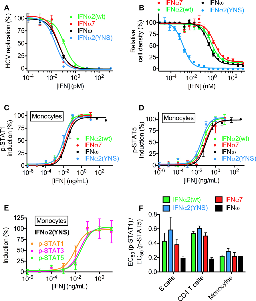
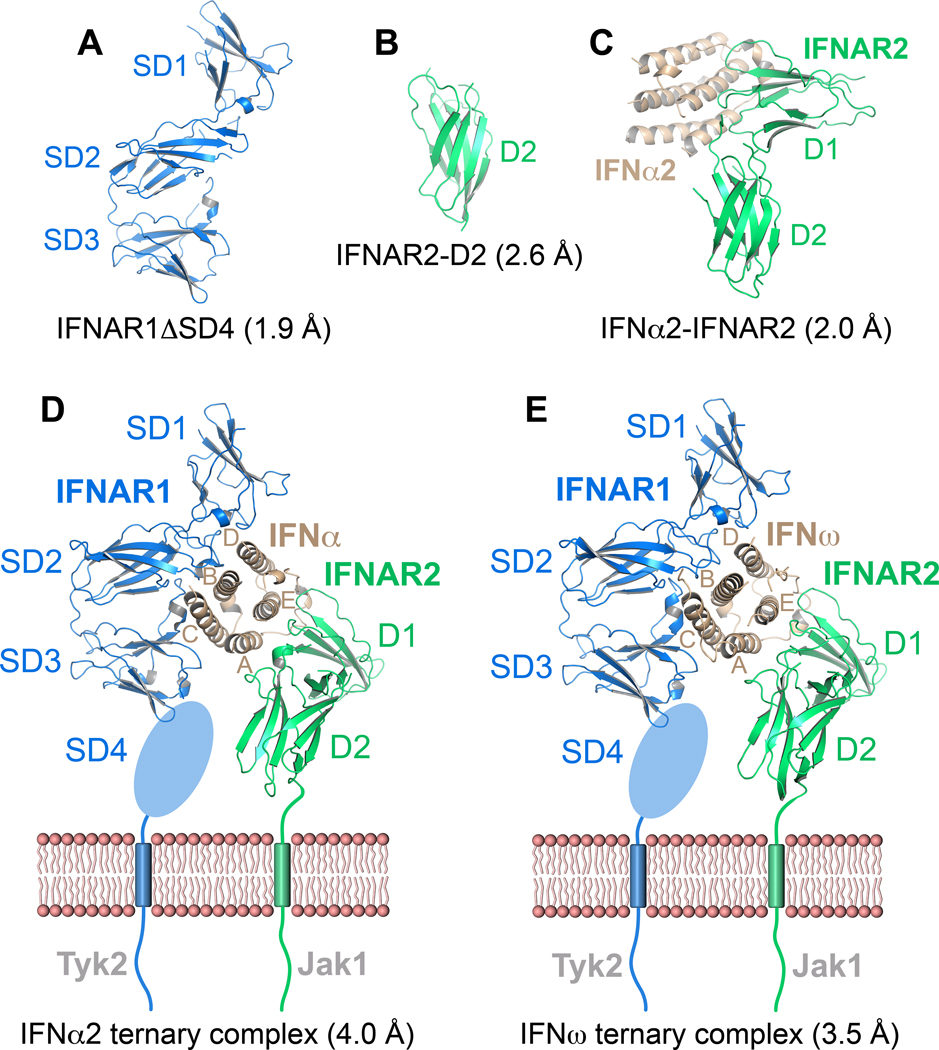
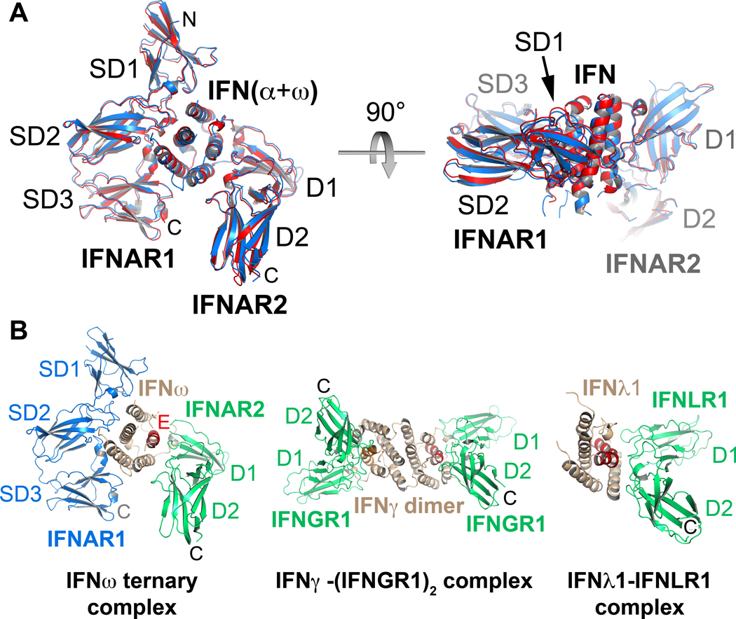
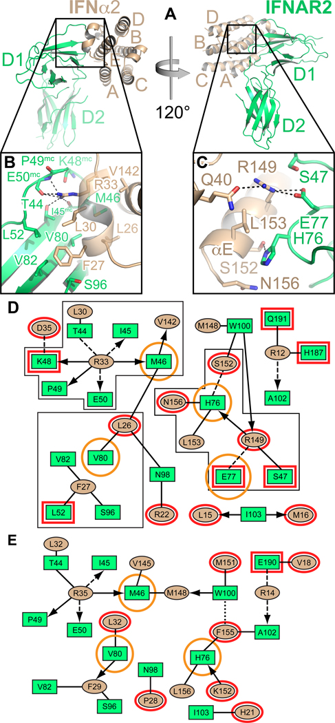
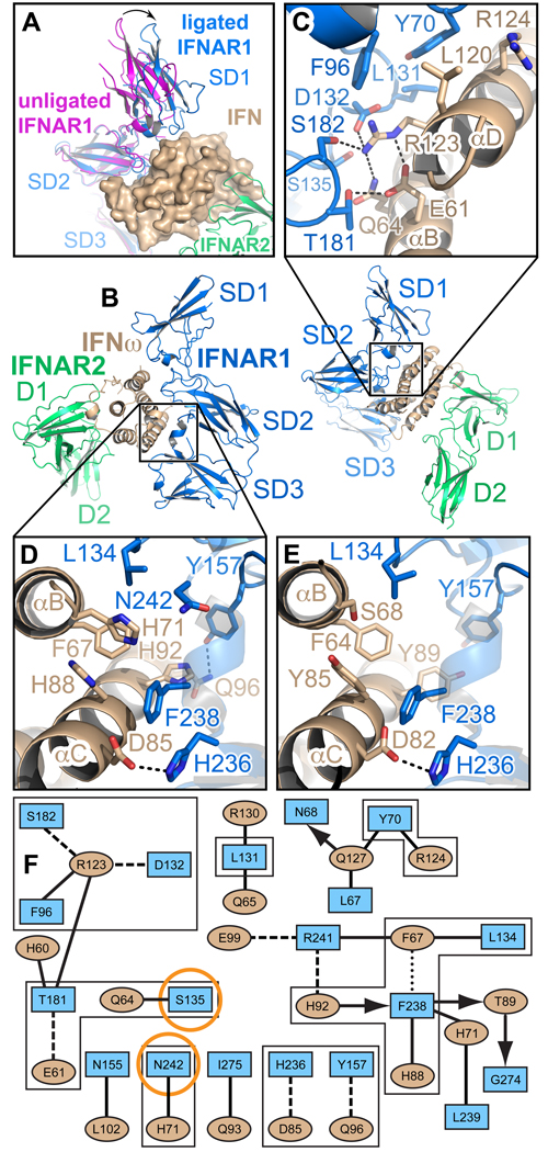
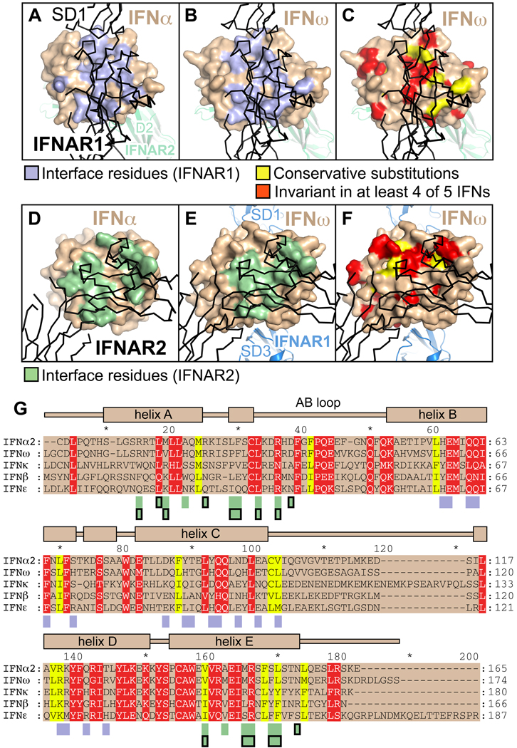
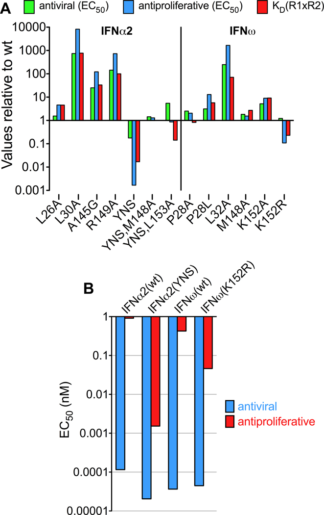
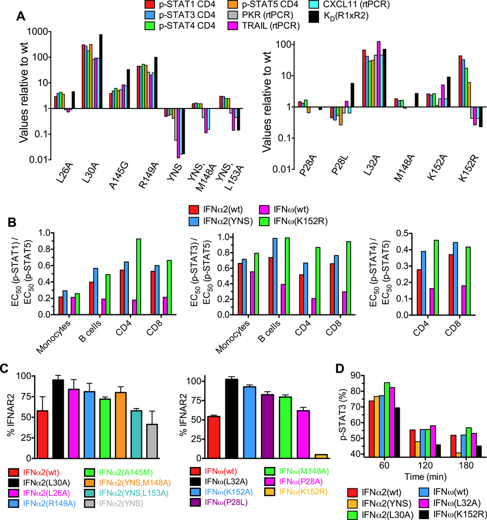
Type I Interferons (IFNs) are important cytokines for innate immunity against viruses and cancer. Sixteen human type I IFN variants signal through the same cell-surface receptors, IFNAR1 and IFNAR2, yet they can evoke markedly different physiological effects. The crystal structures of two human type I IFN ternary signaling complexes containing IFNα2 and IFNω reveal recognition modes and heterotrimeric architectures that are unique among the cytokine receptor superfamily but conserved between different type I IFNs. Receptor-ligand cross-reactivity is enabled by conserved receptor-ligand "anchor points" interspersed among ligand-specific interactions that "tune" the relative IFN-binding affinities, in an apparent extracellular "ligand proofreading" mechanism that modulates biological activity. Functional differences between IFNs are linked to their respective receptor recognition chemistries, in concert with a ligand-induced conformational change in IFNAR1, that collectively control signal initiation and complex stability, ultimately regulating differential STAT phosphorylation profiles, receptor internalization rates, and downstream gene expression patterns.


Figures

Figure 1
Figure 1. Differential activities and potencies of type I IFNs
(A) Antiviral dose-response curves of human hepatoma (Huh7.5) cells transfected with genomic hepatitis C virus (HCV) RNA and treated with IFNα2(wt), IFNα2(YNS), IFNω or IFNα7. (B) Antiproliferative dose-response curves of human amniotic epithelial (WISH) cells treated with IFNα2(wt), IFNα2(YNS), IFNω or IFNα7. (C) Dose-response curves for STAT1 phosphorylation in monocytes from human whole blood, as determined by Phospho-Flow analysis. (D) Dose-response curves for STAT5 phosphorylation in monocytes from human whole blood, as determined by Phospho-Flow cytometry analysis. (E) IFNα2(YNS) more potently induces pSTAT1 than pSTAT3 or pSTAT5 in human primary monocytes. (F) Differential signaling properties of IFNα2(wt), IFNα2(YNS), IFNω and IFNα7 as evidenced by different ratios of pSTAT1 to pSTAT5 EC50 values in different cell types from human whole blood. See also Figure S1.
Figure 2
Figure 2. Crystal structures of type I IFN receptor components and ligand-receptor complexes
Ribbon representations and designated resolutions of (A) IFNAR1ΔSD4. (B) IFNAR2-D2. (C) The IFNα2(HEQ)-IFNAR2 binary complex (IFNα2(HEQ) brown, IFNAR2 green). (D) Ternary complex of IFNAR1 (blue), IFNAR2 (green) and IFNα2(YNS). (E) Ternary complex of IFNAR1 (blue), IFNAR2 (green) and IFNω. The membrane-proximal SD4 domain of IFNAR1 is depicted as an oval. See also Table S1 and Figure S2.
Figure 3
Figure 3. Similar architectures of type I IFN complexes are distinct from type II and type III IFN receptor complexes
(A) The IFN molecules of the IFNω and IFNα2(YNS) ternary complexes were superimposed and are shown in side view and top view. The RMSD for the overall superposition of both structures is 0.9 Å. (B) The IFNω ternary complex is shown side-by-side with the IFNγ-(IFNGR1)2 complex (PDB accession code: 1FG9), and the IFNλ-IFNLR1 complex (PDB accession code: 3OG6). (N, C: amino- and carboxy-termini. SD1–SD4: subdomains of IFNAR1; D1, D2: N- and C-terminal domain of IFNAR2. See also Figure S3B.
Figure 4
Figure 4. Specificity and cross-reactivity determinants between IFN-IFNAR2
(A) Two different views of the IFNα2-IFNAR2 binary complex. Helices of IFNα2 are labeled A–E. (B) Hotspot residues Leu30α2 and Arg33α2 of IFNα2 and their environment in the interface with IFNAR2. Hydrogen bonds are shown as dashed lines. (C) Close-up view of Arg149IFN, Glu77R2 and His76R2 and their environment. Hydrogen bonds and salt bridges are depicted as dashed lines. (D) 2D interaction map of the IFNα2-IFNAR2 interface. Amino acids are depicted as nodes in the interaction maps (rectangles: IFNAR2; ellipses: IFN). Interactions between side chains are represented by lines, interactions between side chains and backbone are depicted as arrows pointing toward the backbone. Van-der-Waals interactions and hydrophobic contacts are shown as solid lines, H-bonds or electrostatic interactions as dashed lines and aromatic interactions as dotted lines. Residues shown in panels B and C are bordered with a black line. Structural differences between the IFNα-IFNAR2 and the IFNω-IFNAR2 interfaces are highlighted in red. IFNAR2 residues that, when mutated, differentially affect IFNα and IFNω binding, are encircled in orange. (E) 2D interaction map of the IFNω-IFNAR2 interface. See also Figure S2 and Table S2.
Figure 5
Figure 5. Ligand-induced domain movement in IFNAR1 and the IFN-IFNAR1 interfaces
(A) Domain movement in IFNAR1 upon IFN binding. Unliganded IFNAR1ΔSD4 (magenta) was superimposed onto subdomains 2 (SD2) and 3 (SD3) of IFNAR1 (blue) in the IFNω ternary complex. The difference in the position of the SD1 domain is depicted as an arrow. The ligand, IFNω, is shown with its molecular surface. See also Figure S3, and http://proteopedia.org/w/Journal:Cell:1. (B) Two different views of the IFNω ternary complex. SD1–SD3: subdomains of IFNAR1; D1 and D2: subdomains of IFNAR2. (C) Environment of the hotspot residues Tyr70R1 and Arg123ω in the IFNAR1-IFNω interface. Dashed lines symbolize hydrogen bonds and salt bridges. (D) Hydrophobic and aromatic interactions between Leu134R1 and the hotspot residue Phe238R1 in IFNAR1 and Phe67 in IFNω. (E) The same region as in (D) in the IFNα2-IFNAR1 interface. Hydrogen bonds in the close-up views are depicted as dashed lines. (F) Interaction map of the IFNω-IFNAR1 interface in the IFNω ternary complex. Amino acids are depicted as nodes in the interaction map (rectangles: IFNAR1; ellipses: IFNω) as used in Figure 3. Residues shown in panels C, D and E are bordered with a black line. IFNAR1 residues that, when mutated, differentially affect IFNα and IFNω binding, are encircled in orange.
Figure 6
Figure 6. Conservation of residues in the ligand-receptor interfaces
(A–F) Residues on the surface of IFNω and IFNα involved in the interaction with IFNAR1 (panel A: IFNα ternary complex, panel B: IFNω ternary complex) and IFNAR2 (panel D: IFNα binary complex, panel E: IFNω ternary complex) are colored lightblue and green, respectively. Surface residues on IFNω conserved between IFNs are shown in panels C and F. Physico-chemically conserved amino acids are colored yellow, residues that are invariant in at least four of five IFNs (IFNα2, IFNβ, IFNε, IFNκ and IFNω) are shown in red. (G) Sequence alignment of human IFNs. Conserved and invariant residues are colored as in panels C and F. Interacting residues are denoted by rectangles below the alignment, colored according to panels A, B, D and E. Rectangles outlined in black mark interacting residues in the IFNα2 binary complex. The secondary structural elements of IFNω are depicted on top of the alignment.
Figure 7
Figure 7. Correlation between complex stability and functional activity
(A) Antiviral and antiproliferative activity of IFNα2 and IFNω mutants relative to IFNα2(wt) and IFNω(wt), respectively. As a measure of complex stability, the product of the affinities toward IFNAR1 and IFNAR2 was calculated and divided by the value of the respective wildtype protein. (B) Direct comparison of the antiviral and antiproliferative activity (EC50 values) of the high-affinity mutants IFNα2(YNS) and IFNω(K152R) and the corresponding wildtype proteins. See also Figure S5.
Figure 8
Figure 8. Relationship between STAT phosphorylation, gene expression, and receptor downregulation to IFN mutant binding affinities
(A) Complex stabilities and induction of STAT phosphorylation in CD4 T cells and gene expression [Protein kinase R (PKR), Tumor necrosis factor-related apoptosis-inducing ligand (TRAIL), chemokine CXCL11] by IFNα2 and IFNω mutants relative to wildtype IFNα2 and IFNω, respectively. (B) Different EC50(p-STAT) ratios for IFNα2(YNS), IFNω(K152R), IFNα2(wt) and IFNω(wt) in different cell subsets of whole blood from human donors. IFNα2(YNS) and IFNω(K152R) show the same trend of ratio deviations from the wildtype proteins. (C) Expression levels of IFNAR2 on the surface of B cell lymphoma (Ramos) cells 5 min after stimulation with IFNα2 and IFNω proteins. (D) Time course of decrease of p-STAT3 activation induced by different IFNα and IFNω proteins. See also Figures S6 and S7.

Comment in

Similar articles

Cited by

References

    1. Adams PD, Afonine PV, Bunkoczi G, Chen VB, Davis IW, Echols N, Headd JJ, Hung LW, Kapral GJ, Grosse-Kunstleve RW, et al. PHENIX: a comprehensive Python-based system for macromolecular structure solution. Acta Crystallogr D Biol Crystallogr. 2010;66:213–221. - PMC - PubMed
    1. Bleicher L, de Moura PR, Watanabe L, Colau D, Dumoutier L, Renauld JC, Polikarpov I. Crystal structure of the IL-22/IL-22R1 complex and its implications for the IL-22 signaling mechanism. FEBS Lett. 2008;582:2985–2992. - PubMed
    1. Bogdan C. The function of type I interferons in antimicrobial immunity. Curr Opin Immunol. 2000;12:419–424. - PubMed
    1. Borden EC, Sen GC, Uze G, Silverman RH, Ransohoff RM, Foster GR, Stark GR. Interferons at age 50: past, current and future impact on biomedicine. Nat Rev Drug Discov. 2007;6:975–990. - PMC - PubMed
    1. Cajean-Feroldi C, Nosal F, Nardeux PC, Gallet X, Guymarho J, Baychelier F, Sempe P, Tovey MG, Escary JL, Eid P. Identification of residues of the IFNAR1 chain of the type I human interferon receptor critical for ligand binding and biological activity. Biochemistry. 2004;43:12498–12512. - PubMed

Publication types