. 2011 Nov 15;418(2):184-96.
doi: 10.1016/j.ab.2011.07.024. Epub 2011 Jul 28.

A methodology for simultaneous fluorogenic derivatization and boronate affinity enrichment of 3-nitrotyrosine-containing peptides

Affiliations

A methodology for simultaneous fluorogenic derivatization and boronate affinity enrichment of 3-nitrotyrosine-containing peptides

Elena S Dremina et al. Anal Biochem. .

Abstract

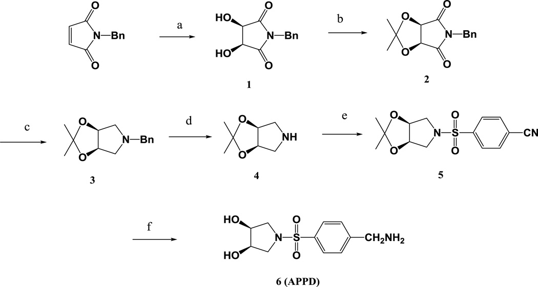
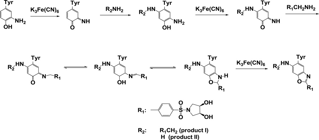
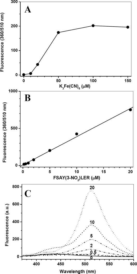
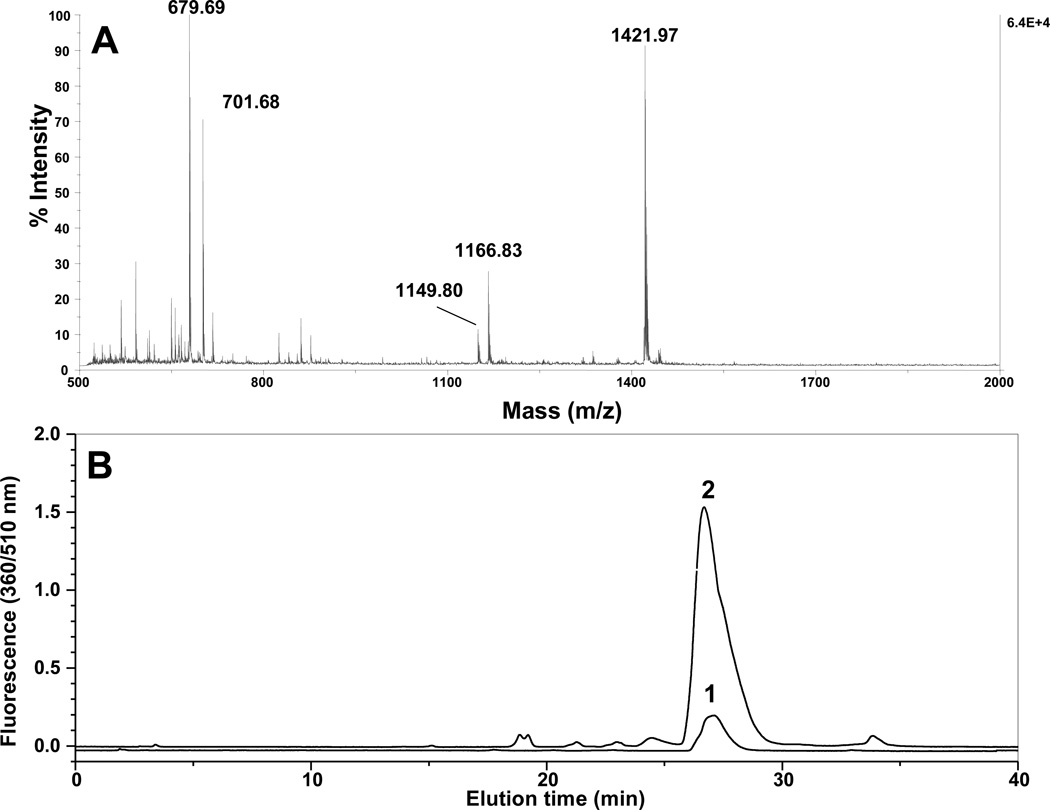
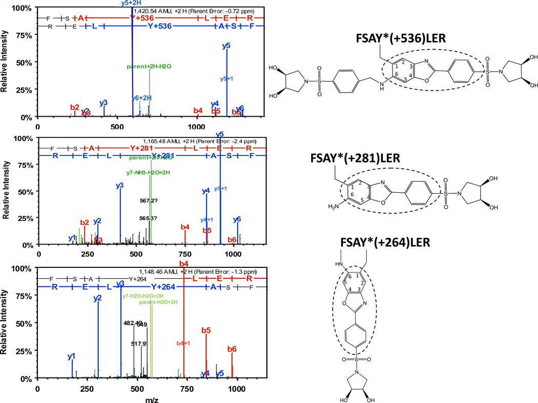
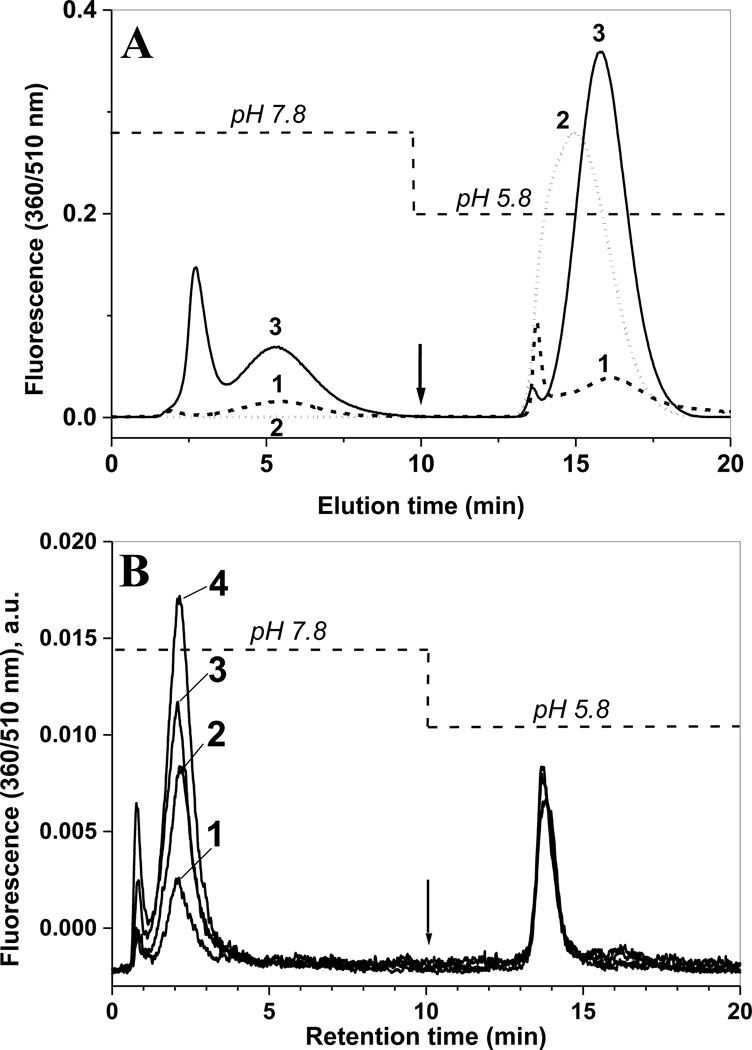
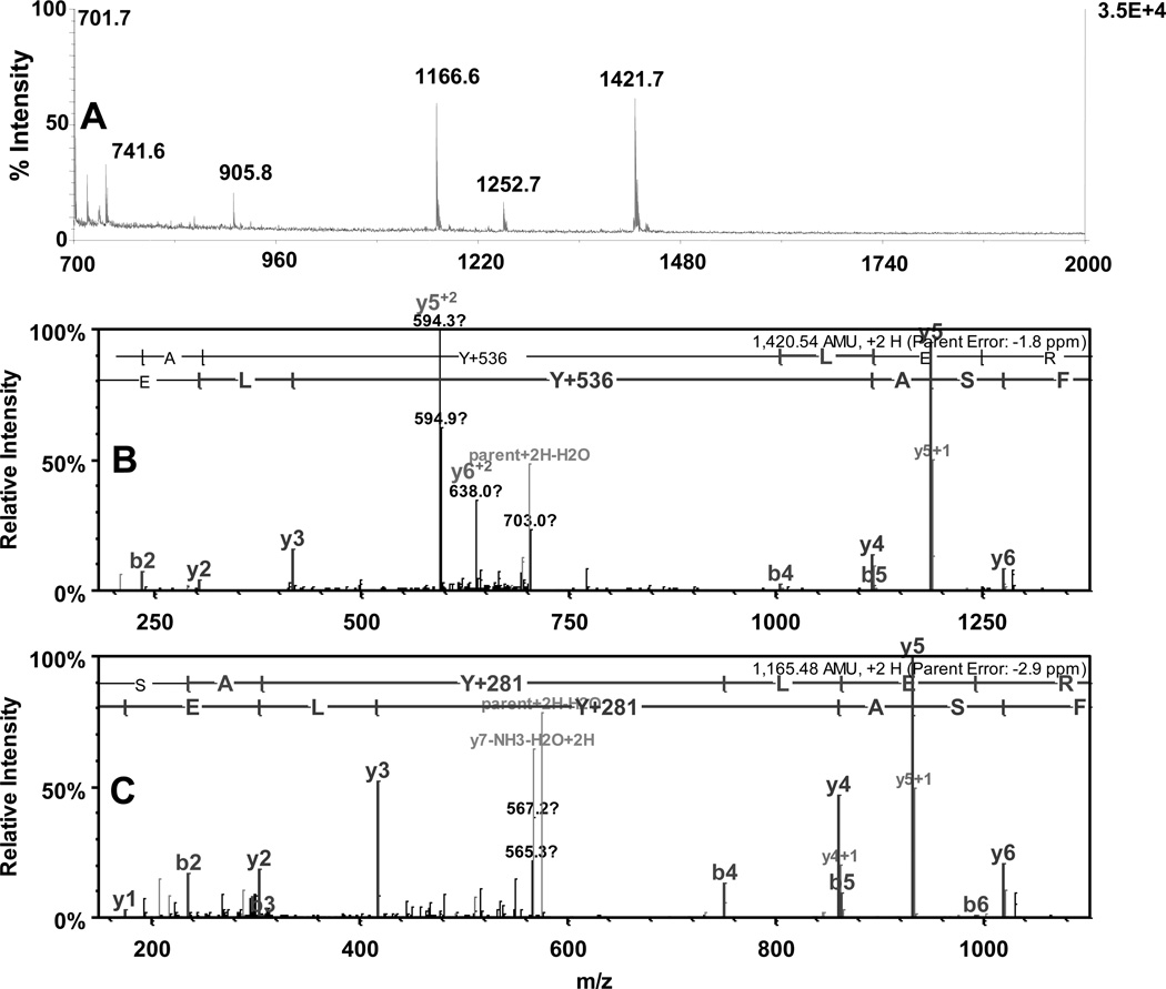
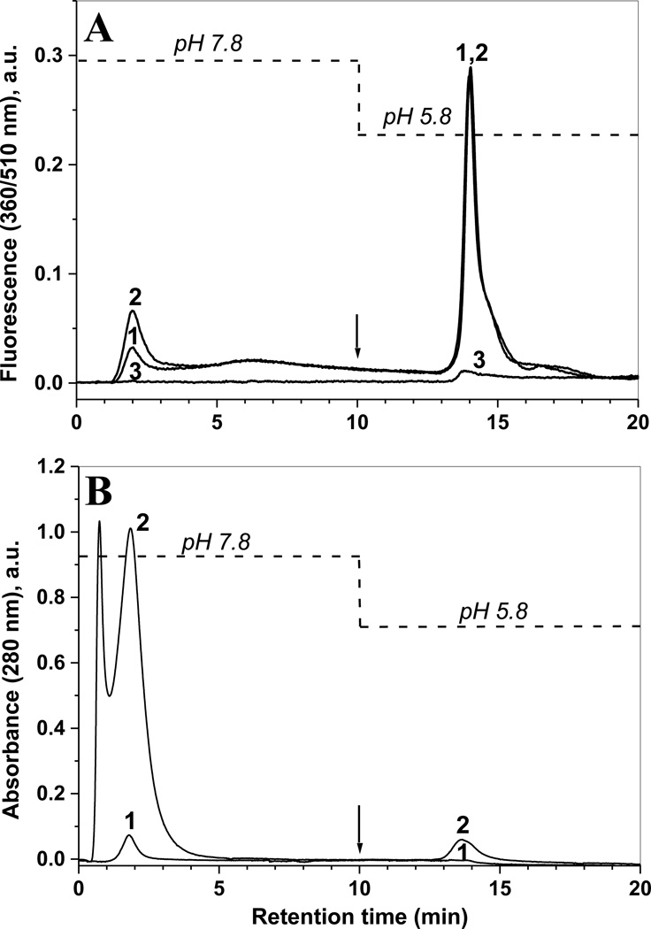
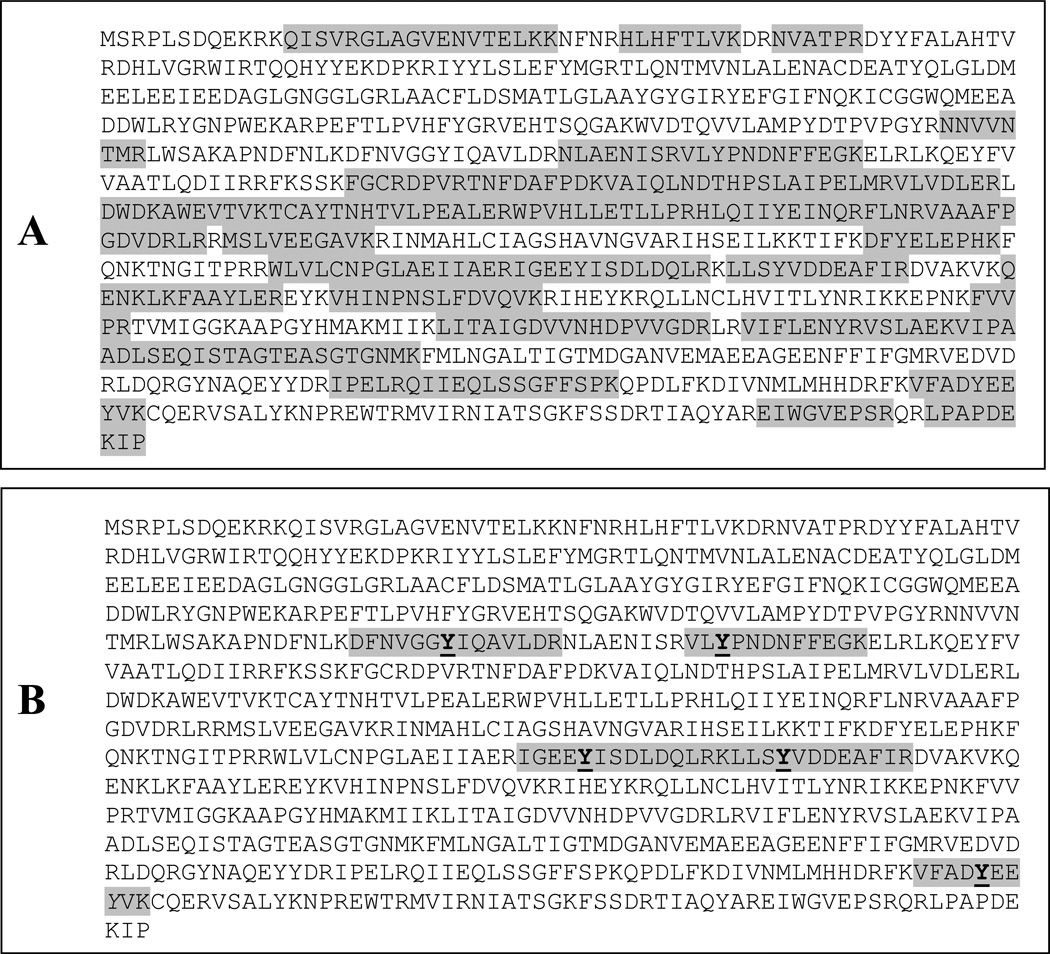
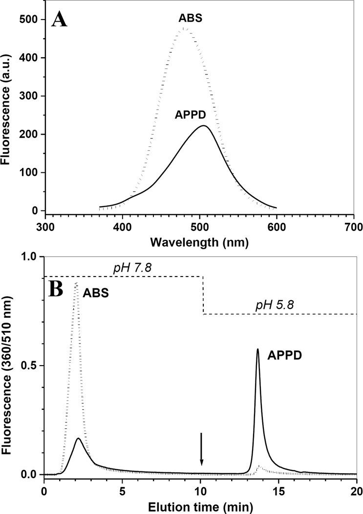
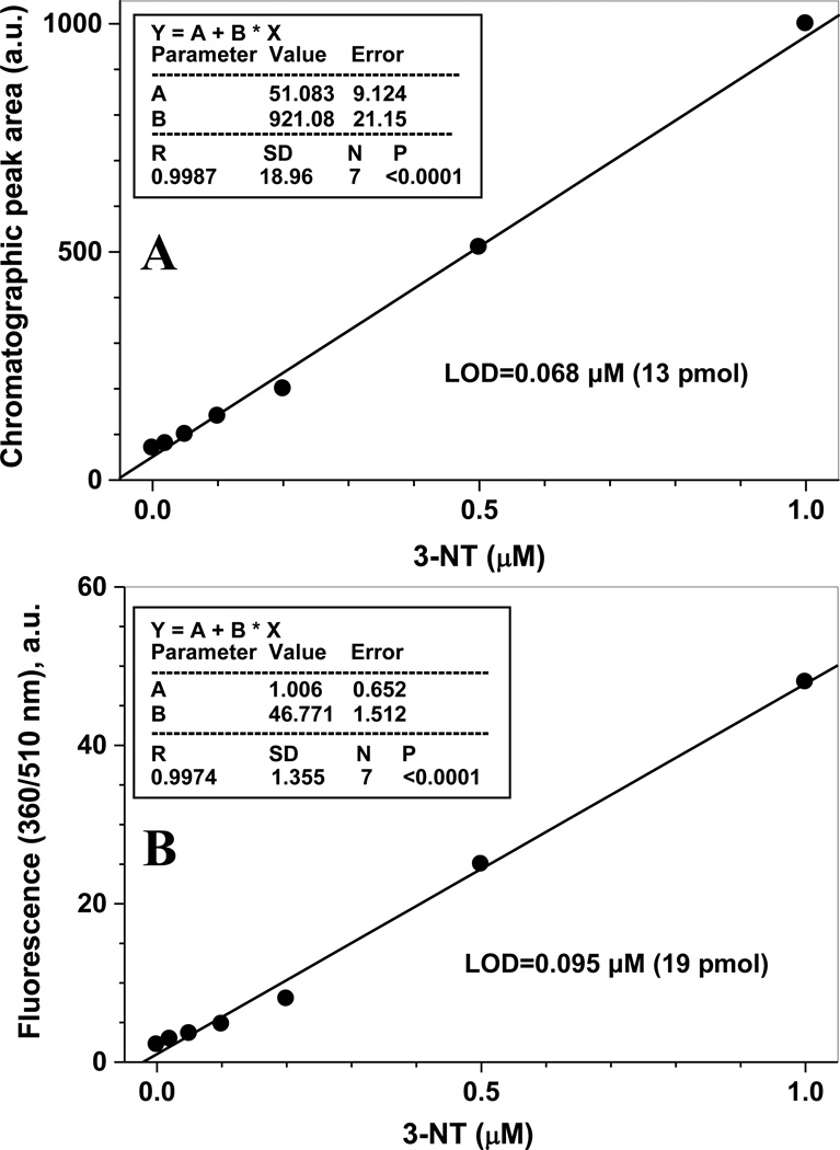
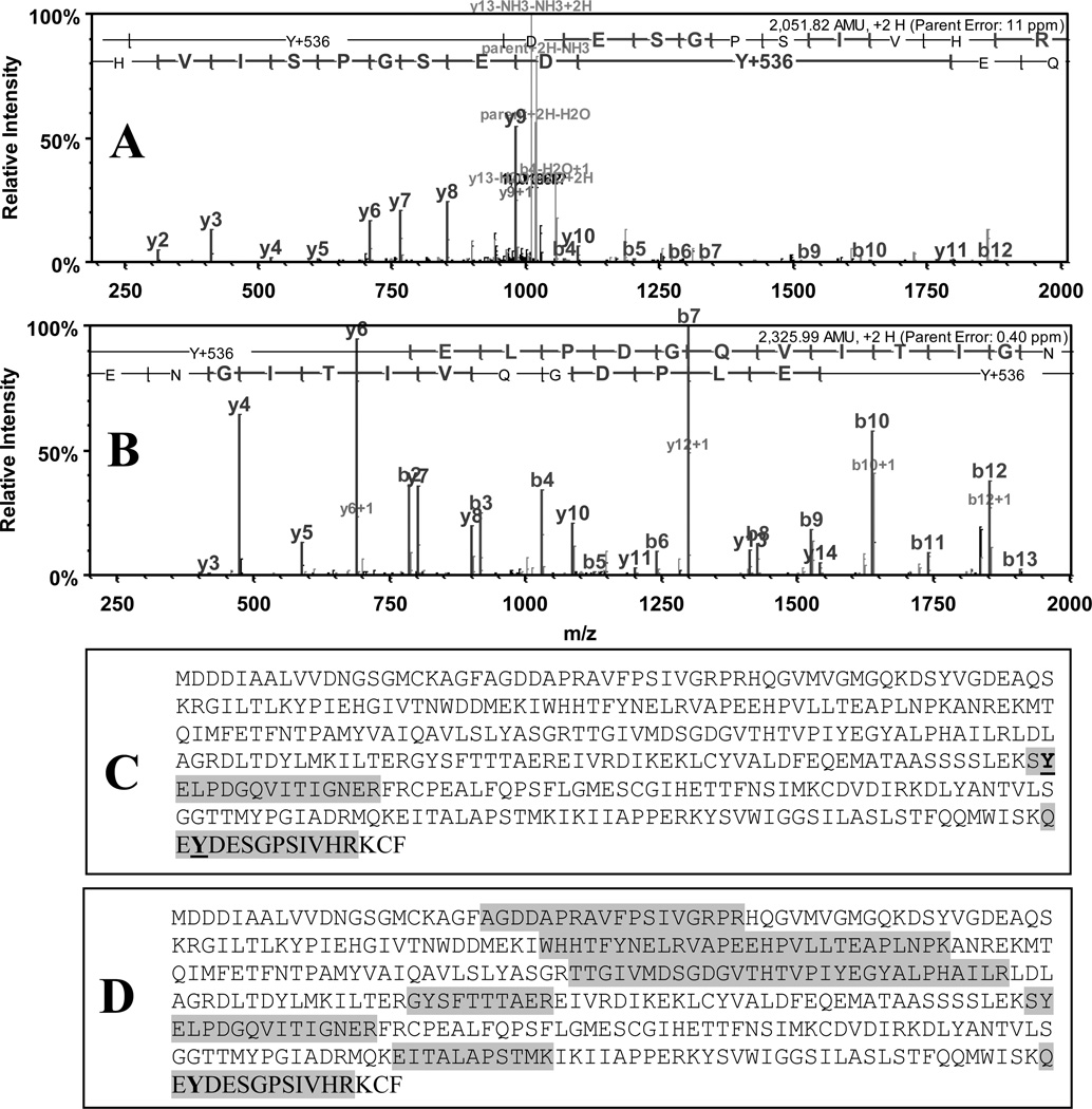
We synthesized and characterized a new tagging reagent, (3R,4S)-1-(4-(aminomethyl)phenylsulfonyl)pyrrolidine-3,4-diol (APPD), for the selective fluorogenic derivatization of 3-nitrotyrosine (3-NT) residues in peptides (after reduction to 3-aminotyrosine) and affinity enrichment. The synthetic 3-NT-containing peptide, FSAY(3-NO(2))LER, was employed as a model for method validation. Furthermore, this derivatization protocol was successfully tested for analysis of 3-NT-containing proteins exposed to peroxynitrite in the total protein lysate of cultured C2C12 cells. The quantitation of 3-NT content in samples was achieved through either fluorescence spectrometry or boronate affinity chromatography with detection by specific fluorescence (excitation and emission wavelengths of 360 and 510 nm, respectively); the respective limits of detection were 95 and 68 nM (19 and 13 pmol total amount) of 3-NT. Importantly, the derivatized peptides show a strong retention on a synthetic boronate affinity column, containing sulfonamide-phenylboronic acid, under mild chromatographic conditions, affording a route to separate the derivatized peptides from large amounts (milligrams) of nonderivatized peptides and to enrich them for fluorescent detection and mass spectrometry (MS) identification. Tandem MS analysis identified chemical structures of peptide 3-NT fluorescent derivatives and revealed that the fluorescent derivatives undergo efficient backbone fragmentations, permitting sequence-specific identification of protein nitration at low concentrations of 3-NT in complex protein mixtures.


Figures

Figure 1
Figure 1
Synthesis of APPD. Reagents and conditions: (a) OsO4, NMO, H2O/acetone (5:1 v/v), 50 °C; (b) 2,2-dimethyloxypropane/TyOH; (c) LiAlH4, THF, 80 °C; (d) H2, Pt/C, MeOH; (e) 4-cyanobenzene-1-sulfonyl chloride, Et3N, MeCN; (f) H2, Pt/C, MeOH, 10% conc. HCl.
Figure 2
Figure 2
Derivatization pathway of peptide 3-AT with APPD.
Figure 3
Figure 3
The dependences of fluorescence intensity after tagging of FSAY(3-NO2)LER with APPD on the concentration of K3Fe(CN)6 (A) or 3-NT peptide (B) and fluorescence spectra at different concentrations of 3-NT (C). A - FSAY(3-NO2)LER (5 µM) was reduced to FSAY(3-NH2)LER and incubated with 10 mM APPD and different concentrations of K3Fe(CN)6 in 0.1 M sodium phosphate buffer (pH 9.0) for 1 h. B and C - Fluorescence intensities and spectra, respectively, at different concentrations of the 3-NT peptide tagged with 10 mM APPD and 100 µM K3Fe(CN)6 as described above. Fluorescence was measured in a 0.5-mL (1-cm) cuvette using excitation and emission wavelengths of 360 and 510 nm, respectively.
Figure 4
Figure 4
Analysis by MALDI-TOF MS (A) and reverse-phase HPLC detected by fluorescence (B) of APPD-tagged FSAY(3-NO2)LER. After reduction of 3-NT to 3-AT the peptide (10 µM) was incubated for 1 h with 10 mM APPD and 20 µM (curve 1) or 100 µM (curve 2) of K3Fe(CN)6 in 0.1 M sodium phosphate buffer (pH 9.0) at room temperature. Chromatograms were detected using excitation and fluorescence wavelengths of 360 and 510 nm, respectively.
Figure 5
Figure 5
ESI-MS/MS identification of different benzoxazole products after nitro-to-amino reduction of FSAY(3-NO2)LER (10 µM) and tagging with 10 mM APPD and 100 µM K3Fe(CN)6. The structures shown in inserts contain different substitutions in position 6 of the original 3-NT ring (the fluorescent 2-phenylbenzoxasole core is circled).
Figure 6
Figure 6
Boronic affinity chromatography of APPD tagged synthetic peptide (A) and of tryptic digest of nitrated rabbit phosphorylase b mixed with proteins from lysate of C2C12 cultured cells (B) detected by fluorescence (excitation and emission wavelengths are 360 and 510 nm, respectively). In panel A, 200 µL of 10 µM FSAY(3-NO2)LER after reduction of 3-NT to 3-AT and incubation with 10 mM APPD and 100 µM K3Fe(CN)6 for 1 h was injected onto phenylboronic column equilibrated with buffer A containing 50 mM NH4HCO3 (pH 7.8) in the absence (1) or presence (2 and 3) of 30% (v/v) acetonitrile. The arrow indicates switching from buffer A to water in the mobile phase without changing the percentage of organic solvent. Trace 2 represents a blank injection (washing run) after the separation performed in the absence of organic solvent (trace 1). Panel B: Boronic affinity separation of tryptic peptides from nitrated Ph-b (1 µg protein containing 100 pmol 3-NT) only (trace 1) and in the presence of C2C12 cell proteins (trace 2 – 0.2, trace 3 – 0.5, and trace 4 – 1 mg total protein) reduced and tagged in the same conditions.
Figure 7
Figure 7
Analysis of fluorescent boronate affinity HPLC fractions (13–18 min. in chromatogram 3, Fig.6A) by MALDI-TOF (A) and capLC-FT-ICR MS (B and C). B and C represent tandem MS spectra for APPD-tagged FSAY*LER ions with mass shifts of Y+536 (m/z 1421.5) and Y+281 (m/z 1166.6), respectively, present in panel A.
Figure 8
Figure 8
Boronate affinity chromatography of model nitrated protein, Ph-b, mixed with high amounts of C2C12 cell lysate protein, through APPD tagging of digested proteins and detected by specific fluorescence (A) or 280-nm absorbance (B). Nitrated Ph-b (5 µg) was analyzed either alone (trace 1) or in the presence of 0.2 mg C2C12 proteins (trace 2); trace 3 represents a reagent control (APPD tagging in the absence of 3-NT). 3-NT containing peptides were reduced to corresponding 3-AT peptides and tagged with 10 mM APPD and 50 µM K3Fe(CN)6 in 0.2 M NH4HCO3 (ABC) buffer at pH 9, bound to boronate affinity column in 0.1 M ABC buffer at pH 7.8 (0–10 min) and eluted with H2O at pH 5.8 (10–20 min) at a flow rate of 1 ml/min. Boronate affinity (13–18 min) and flow-through fractions (1–10 min) were collected for LC-MS analysis.
Figure 9
Figure 9
Sequence coverage for Ph-b in flow-through (A) and boronate affinity (B) fractions collected during chromatographic run shown in Fig.8 (trace 2). In panel B, APPD derivatization products detected in the sample (Y+536, Y+ 264, and Y+281) are shown in bold, italic and underlined font, respectively.
Figure 10
Figure 10
Comparison of fluorescence spectra (A) and boronate affinity chromatographic runs detected by fluorescence (B) of nitrated C2C12 cultured cell protein digests (250 µg total protein containing 500 nmol 3-NT), reduced with 20 mM sodium dithionite and tagged with 10 mM of either APPD (solid lines) or ABS (dotted lines) and 100 µM K3Fe(CN)6 in 100 mM PBS (pH 9.0). Chromatographic conditions were as in the legend to Fig.8.
Figure 11
Figure 11
Validation of the fluorogenic APPD derivatization as quantitative method for peptide 3-NT determination by boronate affinity chromatography with fluorescence detection (A) and by fluorescence spectrometry (B). 3-NT concentration in samples (0–1 µM) was achieved through addition of different amounts of nitrated Ph-b, containing ca. 10 mol 3-NT/mol protein (1 µM 3-NT/2.5 µg Ph-b), to 0.5 mg of C2C12 cell lysate protein. Samples were digested with trypsin, reduced by 10 mm sodium dithionite and derivatized with 2 mM APPD and 20 µM K3Fe(CN)6 in 50 mM ammonium bicarbonate buffer (pH 9.0). Boronate affinity chromatography conditions were as in the legend to Fig. 8.
Figure 12
Figure 12
Representative capLC-FT-ICR tandem MS spectra of APPD-tagged peptides in the protein tryptic digest from peroxynitrite-exposed C2C12 cells, obtained after boronate affinity enrichment of a sample contained 1 nmol 3-NT in 500 µg of protein. A and B represent tandem MS spectra of two peptides, SY*ELPDGQVITIGNER and QEY*DESGPSIVHR (mass shift on both of +536 relative to Tyr), from the sequence of mouse cytoplasmic actin 1 (gene name: actb). Protein sequence coverage is shown in panel C; for comparison, panel D shows sequence coverage for this protein observed without APPD tagging and boronate affinity enrichment.

References

    1. Turko IV, Murad F. Protein nitration in cardiovascular diseases. Pharmacol. Rev. 2002;54:619–634. - PubMed
    1. Reynolds MR, Berry RW, Binder L. Nitration in neurodegeneration: deciphering the “Hows” “nYs”. Biochemistry. 2007;46:7325–7336. - PubMed
    1. Ischiropoulos H. Biological selectivity and functional aspects of protein tyrosine nitration. Biochem. Biophys. Res. Commun. 2002;305:776–783. - PubMed
    1. Radi R. Nitric oxide, oxidants, and protein tyrosine nitration. Proc. Natl. Acad. Sci. USA. 2004;101:4003–4008. - PMC - PubMed
    1. Pacher P, Beckman JS, Liaudet L. Nitric oxide and peroxynitrite in health and disease. Physiol. Rev. 2007;87:315–424. - PMC - PubMed

Publication types

MeSH terms