. 2011 Dec 1;82(11):1562-71.
doi: 10.1016/j.bcp.2011.08.002. Epub 2011 Aug 17.

In vitro and in vivo modulation of ABCG2 by functionalized aurones and structurally related analogs

Affiliations

In vitro and in vivo modulation of ABCG2 by functionalized aurones and structurally related analogs

Hong-May Sim et al. Biochem Pharmacol. .

Abstract

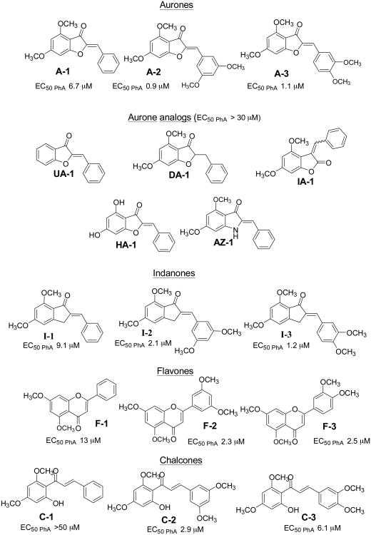
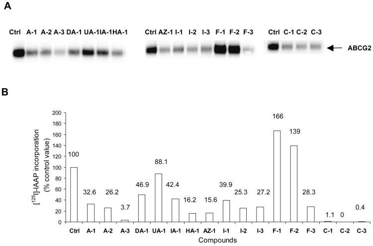
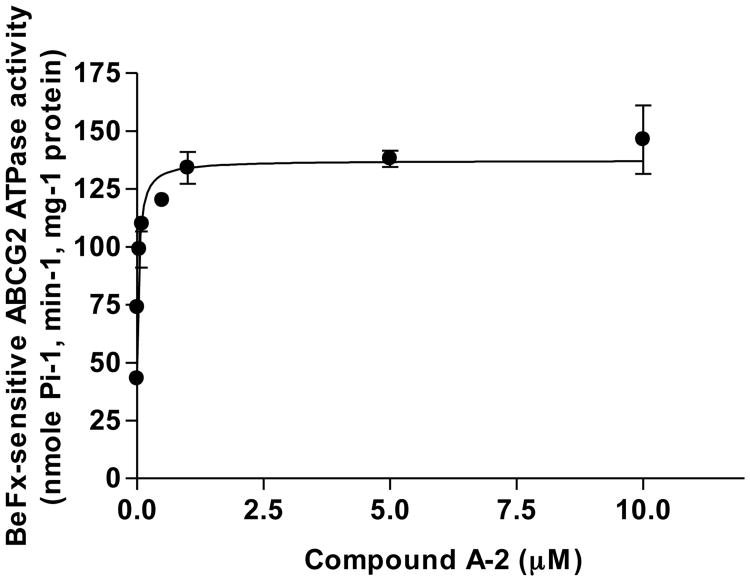
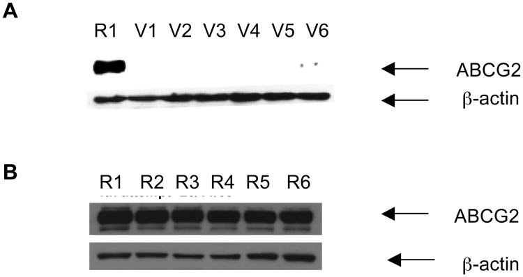
Over-expression of ABCG2 is linked to multidrug resistance in cancer chemotherapy. We have previously shown that functionalized aurones effectively reduced the efflux of pheophorbide A (an ABCG2 substrate) from ABCG2 over-expressing MDA-MB-231/R ("R") cells. In the present report, we investigated the functional relevance of this observation and the mechanisms by which it occurs. Aurones and related analogs were investigated for re-sensitization of R cells to mitoxantrone (MX, a chemotherapeutic substrate of ABCG2) in cell-based assays, accumulation of intracellular MX by cell cytometry, interaction with ABCG2 by biochemical assays and in vivo efficacy in MX resistant nude mice xenografts. We found that methoxylated aurones interacted directly with ABCG2 to inhibit efflux activity, possibly by competing for occupancy of one of the substrate binding sites on ABCG2. The present evidence suggests that they are not transported by ABCG2 although they stimulate ABCG2-ATPase activity. Alteration of ABCG2 protein expression was also discounted. One member was found to re-sensitize R cells to MX in both in vitro and in vivo settings. Our study identified methoxylated aurones as promising compounds associated with low toxicities and potent modulatory effects on the ABCG2 efflux protein. Thus, they warrant further scrutiny as lead templates for development as reversal agents of multidrug resistance.


Figures

Figure 1
Figure 1. Chemical structures of test compounds studied in this work
The EC50 value of each compound for Pheophorbide A accumulation in ABCG2 over-expressing MDA-MB-231/R cells is given at the bottom of the structure.
Figure 2
Figure 2. Accumulation of mitoxantrone in MDA-MB-231/R cells in the absence or presence of 5 μM test compounds
MDA-MB-231/R cells (106cells/ml) were incubated in the absence (control) or presence of 5μM test compound in serum-free RPMI medium for 15 min, 37°C, followed by addition of MX (3 μM) for 30 min. The cells were then washed with ice-cold PBS and resuspended in PBS for the determination of MX on the flow cytometer as described in Methods. 10μM FTC was used a positive control. The MX accumulation in MDA-MB-231/R cells was calculated as a percentage of control (0.1% DMSO) as described previously (Sim et al., 2008). Data points are expressed as mean and error bars represent SD for n = 3 - 4 independent determinations. Statistical difference (*p < 0.05) between MX accumulation in control and treated groups were analyzed using one-way ANOVA analysis followed by Dunnett post-hoc test.
Figure 3
Figure 3. The effect of compound A-2 on the ATPase activity of ABCG2
The ABCG2-expressing High-Five cells crude membrane proteins (10 μg membrane protein/100 μl) was incubated with increasing concentrations of A-2 in the absence or presence of beryllium fluoride in ATPase assay buffer for 3-5 min at 37°C. The reaction was initiated by the addition of 5 mM ATP. After 20 min at 37°C, 2.5% SDS solution was added to terminate the reaction. ABCG2-specific ATPase activity was recorded as BeFx-sensitive ATPase activity as described previously (Shukla et al., 2006; Shukla et al., 2009). The concentration required for 50% stimulation with A-2 was 0.037 ± 0.013 μM (SD, n = 3 independent determinations).
Figure 4
Figure 4. Effect of aurones and their analogs on the photoaffinity labelling of ABCG2 with [125I]-IAAP
Panel A: Autoradiogram showing the photoaffinity labeling of ABCG2 with [125I]-IAAP in the absence or presence of 5μM test compounds. ABCG2-containing crude membranes (50μg protein/ml) were incubated with 5 μM of test compound or FTC for 10 minutes at room temperature in 50 mM Tris-HCl (pH 7.5), after which 3-6 nM [125I] IAAP was added. The samples were incubated for an additional 5 min under subdued light, then exposed to ultravolet (365nm) light for 10 min at room temperature. The labeled ABCG2 was immunoprecipitated using the BXP-21 antibody and processed as described previously (Shukla et al., 2006). The radioactivity incorporated into ABCG2 was determined by exposing the gel to an X-ray film at −70°C and quantified using a phosphoimager and ImageQuaNT software. Panel B: %[125I]-IAAP incorporation = [125I]-IAAP signals obtained in the presence of test compound (5 μM) – [125I]-IAAP labelling obtained in the presence of FTC (5 μM). Values were average from two independent experiments.
Figures 5
Figures 5. Western blot analyses showing the effect of compound A-2 (alone or in combination with mitoxantrone) on ABCG2 expression in MDA-MB-231/V (A) and R cells (B)
Cells were treated under different conditions for 72 hours after which they were trypsinized and 15 μg protein of R and V cell lysates were subjected to electrophoresis on 7.5% SDS-PAGE and transferred to nitrocellulose membranes for overnight blocking at room temperature. The blots were probed with anti-ABCG2 (BXP-21), followed by horseradish peroxidase-conjugated anti-mouse secondary antibody as described previously (Sim et al., 2008). β-Actin was used as a positive control in the Western blot analyses. Blots presented are representative of those from three independent determinations. Lanes marked R or V refer to lysates derived from R or V cells respectively. In panel A, lane R1 and lane V1, control R and V cells in 1% DMSO; lane V2 and V3, V cells exposed to A-2 at 0.5 μM and 1 μM; lane V4, V cells exposed to MX at 0.01 μM; and lane V5 and V6, V cells exposed to A-2 (0.5μM) + MX (0.01 μM) and A-2 (1 μM) + MX (0.01 μM). In panel B, lane R2 and R3, R cells exposed to A-2 at 0.5μM and 1 μM; lane R4, R cells exposed to MX at 0.1 μM; and lane R5 and R6, R cells exposed to A-2 (0.5 μM) + MX (0.1 μM) and A-2 (1 μM) + MX (0.1 μM).

References

    1. Ambudkar SV, Dey S, Hrycyna CA, Ramachandra M, Pastan I, Gottesman MM. Biochemical, cellular, and pharmacological aspects of the multidrug transporter. Annu Rev Pharmacol Toxicol. 1999;39:361–98. - PubMed
    1. Perez-Tomas R. Multidrug resistance: retrospect and prospects in anti-cancer drug treatment. Curr Med Chem. 2006;13:1859–76. - PubMed
    1. Szakacs G, Paterson JK, Ludwig JA, Booth-Genthe C, Gottesman MM. Targeting multidrug resistance in cancer. Nat Rev Drug Discov. 2006;5:219–34. - PubMed
    1. Boumendjel A, Florin A, Boutonnat J. Reversal agents of multidrug resistance mediated by multidrug resistance-associated proteins (MRPs) In: Boumendjel A, Boutonnat J, Robert J, editors. ABC Transporters and Multidrug Resistance. Hoboken, New Jersey: John Wiley & Sons, Inc; 2009. pp. 261–80.
    1. Lee C. Reversing agents for ATP-binding cassette drug transporters. Methods Mol Biol. 2010;596:325–40. - PubMed

Publication types

MeSH terms