. 2011 Nov;176(2):229-37.
doi: 10.1016/j.jsb.2011.08.002. Epub 2011 Aug 10.

A novel structural mechanism for redox regulation of uridine phosphorylase 2 activity

Affiliations

A novel structural mechanism for redox regulation of uridine phosphorylase 2 activity

Tarmo P Roosild et al. J Struct Biol. 2011 Nov.

Abstract

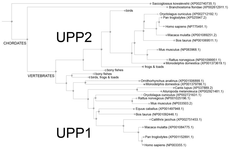
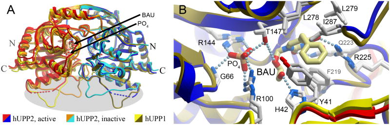
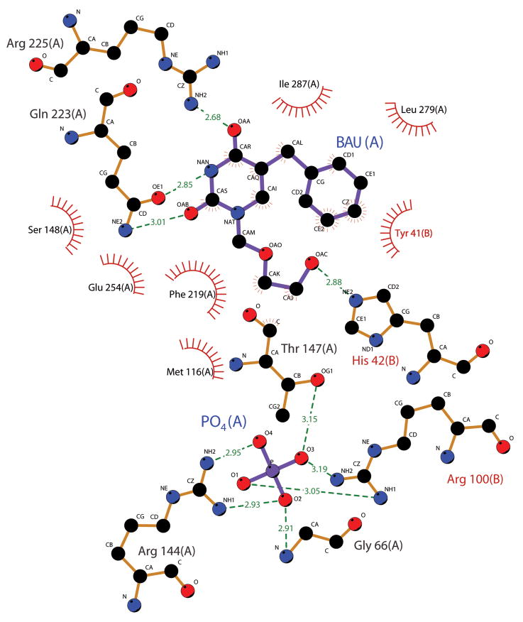
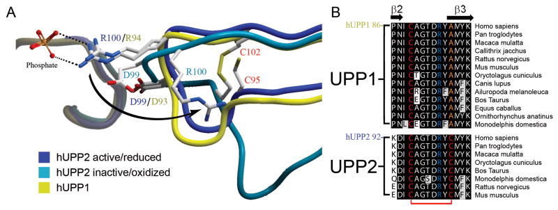
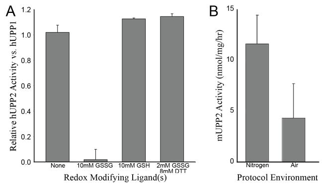
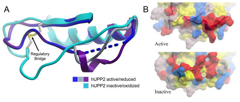
Uridine phosphorylase (UPP) catalyzes the reversible conversion of uridine to uracil and ribose-1-phosphate and plays an important pharmacological role in activating fluoropyrimidine nucleoside chemotherapeutic agents such as 5-fluorouracil and capecitabine. Most vertebrate animals, including humans, possess two homologs of this enzyme (UPP1 & UPP2), of which UPP1 has been more thoroughly studied and is better characterized. Here, we report two crystallographic structures of human UPP2 (hUPP2) in distinctly active and inactive conformations. These structures reveal that a conditional intramolecular disulfide bridge can form within the protein that dislocates a critical phosphate-coordinating arginine residue (R100) away from the active site, disabling the enzyme. In vitro activity measurements on both recombinant hUPP2 and native mouse UPP2 confirm the redox sensitivity of this enzyme, in contrast to UPP1. Sequence analysis shows that this feature is conserved among UPP2 homologs and lacking in all UPP1 proteins due to the absence of a necessary cysteine residue. The state of the disulfide bridge has further structural consequences for one face of the enzyme that suggest UPP2 may have additional functions in sensing and initiating cellular responses to oxidative stress. The molecular details surrounding these dynamic aspects of hUPP2 structure and regulation provide new insights as to how novel inhibitors of this protein may be developed with improved specificity and affinity. As uridine is emerging as a promising protective compound in neuro-degenerative diseases, including Alzheimer's and Parkinson's, understanding the regulatory mechanisms underlying UPP control of uridine concentration is key to improving clinical outcomes in these illnesses.


Figures

Fig. 1
Fig. 1
Phylogenetic tree of all known vertebrate UPP sequences. The presence of two homologues of UPP appears to have arisen early in vertebrates and is retained in most mammals, though some individual animals seem to have subsequently lost UPP2.
Fig. 2
Fig. 2
Comparison of the structure of hUPP2 with hUPP1. (A) Overlay of two alternate structures of hUPP2 bound to BAU with the structure of hUPP1 bound to BAU (PDB ID 3EUF) reveals the strict retention of both overall fold and all secondary structural elements. The position of the inhibitor and binding site for inorganic phosphate proximate to the dimer interface are indicated. For this figure, the right-side monomers of the dimeric enzymes (blue, turquoise, gold) were least-squares aligned producing slightly greater deviations in the backbone traces of the partnering chains, consistent with inherent interdomain flexibility within these proteins (Roosild et al., 2009; Roosild and Castronovo, 2010). However, substantial variation in the main chain conformation is limited to a single, surface-exposed loop on one face of the enzyme (grey highlight). (B) Comparison of the active site structures of hUPP2 and hUPP1 illustrates strict retention of both the identity and positioning of every residue that contacts the enzymes’ substrates. Amino acid labels correspond to the hUPP2 primary sequence with the equivalent hUPP1 residue number being six less.
Fig. 3
Fig. 3
Depiction of hUPP2 ligand-interacting residues. Residues forming energetically favorable interactions with either the inhibitor BAU or the phosphate substrate are illustrated using Ligplot (Wallace et al., 1995). All residues shown are strictly conserved in identity and position between hUPP2 and hUPP1.
Fig. 4
Fig. 4
Redox inactivation of hUPP2. (A) Alternate structures of hUPP2 reveal two different conformations for the loop bearing a critical phosphate coordinating residue (R100). In one case, the structure is virtually identical to that seen in hUPP1 and other uridine phosphorylases (blue vs. yellow). However, in the second case, the formation of a disulfide bridge between C95 and C102 (red) contorts the shape of this loop and twists the key arginine residue away from the active site (turquoise). Additionally, an acidic residue (D99) is shifted closer to the phosphate binding site, which is unoccupied in this structure. These structural changes suggest that this is a catalytically incompetent conformation of this enzyme. (B) Alignment of all known mammalian UPP sequences shows that the second of the cysteine residues needed for disulfide bridge formation is invariably conserved among UPP2 proteins, but absent from all UPP1 enzymes.
Fig. 5
Fig. 5
Redox sensitivity of UPP2. (A) The relative activity of recombinant hUPP2, normalized to hUPP1, upon exposure to various redox modifying compounds is graphed. hUPP2 activity is inactivated upon exposure to oxidized glutatione (GSSG) and slightly enhanced by reduced glutathione (GSH). Other reducing agents, such as dithiothreitol (DTT), can protect hUPP2 from GSSG inactivation, suggesting the observed effect is due to its redox properties and not through some other mechanism of molecular inhibition. (B) The measured activity of native mouse UPP2 is graphed, varying the composition of atmosphere used during enzyme isolation from liver homogenates and subsequent activity analysis. These results obtained with native enzyme confirm the oxidation sensitivity of this family of proteins.
Fig. 6
Fig. 6
Long-range structural consequences of the state of the regulatory disulfide bridge. (A) Of six independent protein chains in the asymmetric unit of crystals of active hUPP2, four are too disordered for modeling in part of the loop region preceding the disulfide bridge (blue). The remaining two chains reveal partially helical structure (grey & purple), due to stabilization through protein-protein contacts with crystallographic-symmetry-related domains. These conformations differ substantially from the redox inactivated structure of hUPP2, in which this region of the protein is flat and elongated (turquoise). (B) Surface representation of the same region, colored by polarity and comparing active and inactive structures, illustrates the degree to which this face of the enzyme is altered, depending upon its redox state.

References

    1. Al Safarjalani ON, Rais R, Shi J, Schinazi RF, Naguib FN, et al. Modulation of 5-fluorouracil host-toxicity and chemotherapeutic efficacy against human colon tumors by 5-(phenylthio)acyclouridine, a uridine phosphorylase inhibitor. Cancer Chemother Pharmacol. 2006;58:692–698. - PubMed
    1. Brandes N, Schmitt S, Jakob U. Thiol-based redox switches in eukaryotic proteins. Antioxid Redox Signal. 2009;11:997–1014. - PMC - PubMed
    1. Bu W, Settembre EC, el Kouni MH, Ealick SE. Structural basis for inhibition of Escherichia coli uridine phosphorylase by 5-substituted acyclouridines. Acta Crystallogr D Biol Crystallogr. 2005;61:863–872. - PubMed
    1. Buhrman G, Parker B, Sohn J, Rudolph J, Mattos C. Structural mechanism of oxidative regulation of the phosphatase Cdc25B via an intramolecular disulfide bond. Biochemistry. 2005;44:5307–5316. - PubMed
    1. Burling FT, Kniewel R, Buglino JA, Chadha T, Beckwith A, et al. Structure of Escherichia coli uridine phosphorylase at 2.0 Å. Acta Crystallogr D Biol Crystallogr. 2003;59:73–76. - PubMed

Publication types