. 2011 Sep 9;412(4):612-7.
doi: 10.1016/j.bbrc.2011.08.009. Epub 2011 Aug 11.

Direct evidence that RNA inhibits APOBEC3G ssDNA cytidine deaminase activity

Affiliations

Direct evidence that RNA inhibits APOBEC3G ssDNA cytidine deaminase activity

William M McDougall et al. Biochem Biophys Res Commun. .

Abstract

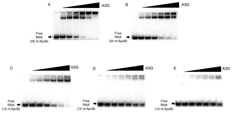
APOBEC3G (A3G) is a deoxycytidine deaminase active on ssDNA substrates. In HIV infected cells A3G interacted with reverse transcription complexes where its activity as a deoxycytidine deaminase led to mutation of the viral genome. A3G not only bound ssDNA, but it also had an intrinsic ability to bind RNA. In many cell types that can support HIV replication, A3G ssDNA deaminase activity was suppressed and the enzyme resided in high molecular mass, ribonucleoprotein complexes associated with cytoplasmic P-bodies and stress granules. Using a defined in vitro system, we show that RNA alone was sufficient to suppress A3G deaminase activity and did so in an RNA concentration-dependent manner. RNAs of diverse sequences and as short as 25nt were effective inhibitors. Native PAGE analyses showed that RNA formed ribonucleoprotein complexes with A3G and in so doing prevented ssDNA substrates from binding to A3G. The data provided direct evidence that A3G binding to cellular RNAs constituted a substantial impediment to the enzyme's ability to interact with ssDNA.


Figures

FIGURE 1
FIGURE 1. A3G assembles multiple complexes on ssDNA and RNA
EMSA using [32P] γ-ATP 5′ end-labeled 41 nt ssDNA or RNA. The concentration of ssDNA in each reaction was 0.6 μM and the concentration of A3G in each lane was 0 μM, 0.22 μM, 0.44 μM, 0.86 μM. Shifts were visualized by phosphorimager scanning densitometry by virtue of their labeled ssDNA content. The gels shown are representative of 4 independent experiments. A. EMSA using 41nt substrate ssDNA containing CCCA (hot spot motif) B. EMSA using 41nt substrate ssDNA containing UUUA. For EMSA using RNAs the concentration of RNA in each reaction was 2.0 μM whereas the concentration of A3G in each lane was 0 μM, 0.03 μM, 0.06 μM, 0.11 μM, 0.22 μM, 0.44 μM, 0.86 μM.. Shifts were visualized by phosphorimager scanning densitometry by virtue of their labeled RNA content. The gels shown are representative of 3 independent experiments. C, D and E are EMSAs using 99nt ApoB RNA, 96nt HIV GAG RNA and 91nt 7SL RNA respectively.
FIGURE 2
FIGURE 2. RNA competes with ssDNA for A3G binding and deaminase activity
EMSA of RNA competition used [32P] γ-ATP 5′ end-labeled 41 nt substrate ssDNA. The concentration of ssDNA in each reaction was 0.6 μM and the concentration of A3G was 1.75 μM. A. The concentration of ApoB 99 RNA, HIV GAG RNA and 7SL (Alu domain) RNA (A, C and E respectively) increased from 0 μM to 60 μM. Each gel is representative of 3 independent experiments. The effect of apoB RNA, HIV GAG RNA and 7SL (Alu domain) RNA (B, D and F respectively) on deaminase activity was assayed in parallel reactions (set up as described above) by poisoned primer extension. The lowest arrow on the left indicates free primer, the middle arrow indicates the primer extension product for deaminated ssDNA (dU) and the upper arrow indicates the primer extension product for unmodified ssDNA (dC). The percent deaminated ssDNA (%dU/dC) was determined by phosphorimager scanning densitometry and calculated as dU divided by (dU + dC) times 100. Each gel is representative of 3 independent experiments.
FIGURE 3
FIGURE 3. Size dependence of A3G assembly on RNA
The concentration of [32P] γ-ATP 5′ end-labeled RNA in each reaction was 2.0 μM, whereas the concentration of A3G varied from 0 μM, 0.03 μM, 0.06 μM, 0.11 μM, 0.22 μM, 0.44 μM, 0.86 μM. Shifts were visualized by phosphorimager scanning densitometry by virtue of their labeled ssDNA content. A. 25nt ApoB RNA, B. 20nt ApoB RNA, C. 15nt ApoB RNA, D. 12nt ApoB RNA, E. 10nt ApoB RNA. The gels are representative of 4 independent experiments.
Figure 4
Figure 4. Size dependence of RNA competition
RNA competition for EMSA used 0.6 μM of [32P] γ-ATP 5′ end-labeled 41 nt substrate ssDNA and 1.75 μM of A3G. The concentration of the 25 nt, 20 nt and 15 nt apoB RNAs (A, B and C respectively) increased from 0 μM to 60 μM. RNA inhibition of deaminase activity was assayed in parallel reactions set up as described by poisoned by poisoned primer extension on 41 nt ssDNA that following incubated with A3G with or without RNA. All gels are representative of 2 independent experiments.

References

    1. Harris RS, Petersen-Mahrt SK, Neuberger MS. RNA editing enzyme APOBEC1 and some of its homologs can act as DNA mutators. Mol Cell. 2002;10:1247–1253. - PubMed
    1. Jarmuz A, Chester A, Bayliss J, Gisbourne J, Dunham I, Scott J, Navaratnam N. An Anthropoid-Specific Locus of Orphan C to U RNA-Editing Enzymes on Chromosome 22. Genomics. 2002;79:285–296. - PubMed
    1. Wedekind JE, Dance GS, Sowden MP, Smith HC. Messenger RNA editing in mammals: new members of the APOBEC family seeking roles in the family business. Trends in genetics : TIG. 2003;19:207–216. - PubMed
    1. Newman EN, Holmes RK, Craig HM, Klein KC, Lingappa JR, Malim MH, Sheehy AM. Antiviral function of APOBEC3G can be dissociated from cytidine deaminase activity. Curr Biol. 2005;15:166–170. - PubMed
    1. Hache G, Liddament MT, Harris RS. The retroviral hypermutation specificity of APOBEC3F and APOBEC3G is governed by the C-terminal DNA cytosine deaminase domain. Journal of Biological Chemistry. 2005;280:10920–10924. - PubMed

Publication types

MeSH terms

LinkOut - more resources