. 2011 Sep;134(Pt 9):2595-609.
doi: 10.1093/brain/awr201. Epub 2011 Aug 19.

FET proteins TAF15 and EWS are selective markers that distinguish FTLD with FUS pathology from amyotrophic lateral sclerosis with FUS mutations

Affiliations

FET proteins TAF15 and EWS are selective markers that distinguish FTLD with FUS pathology from amyotrophic lateral sclerosis with FUS mutations

Manuela Neumann et al. Brain. 2011 Sep.

Abstract

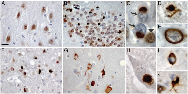
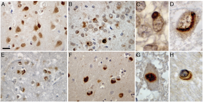
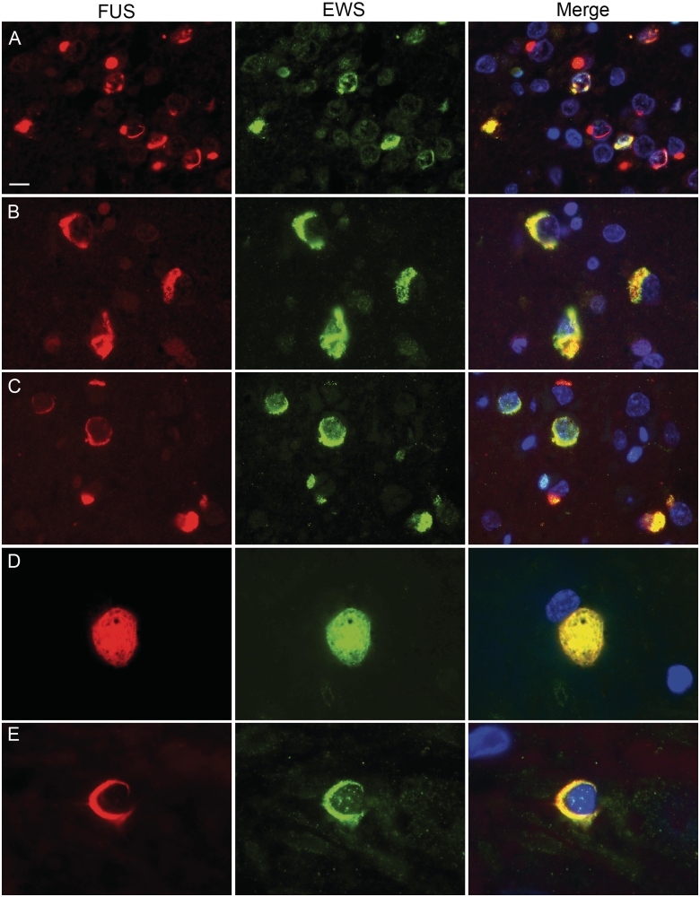
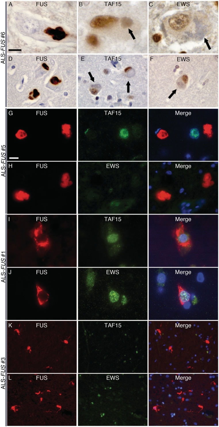
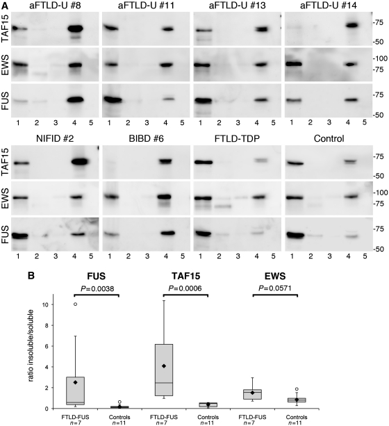
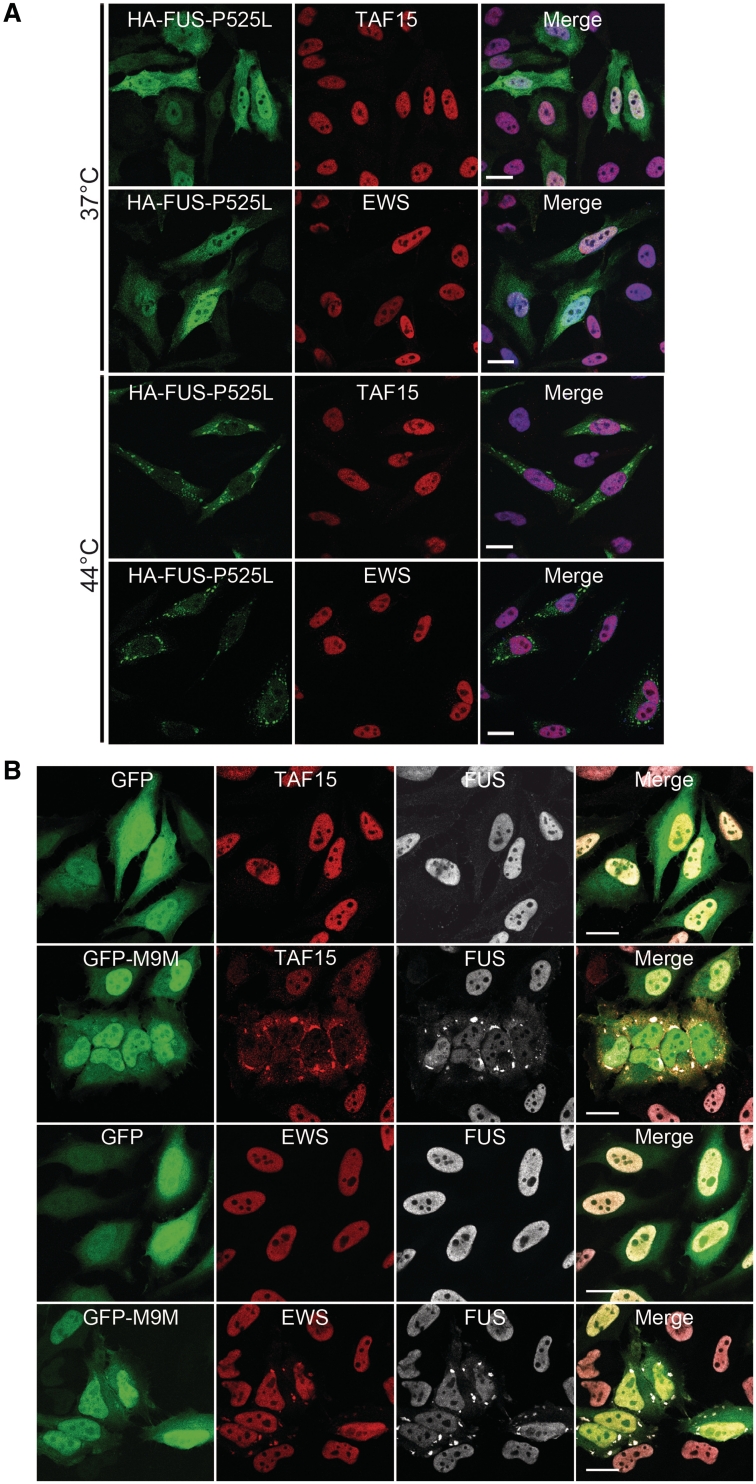
Accumulation of the DNA/RNA binding protein fused in sarcoma as cytoplasmic inclusions in neurons and glial cells is the pathological hallmark of all patients with amyotrophic lateral sclerosis with mutations in FUS as well as in several subtypes of frontotemporal lobar degeneration, which are not associated with FUS mutations. The mechanisms leading to inclusion formation and fused in sarcoma-associated neurodegeneration are only poorly understood. Because fused in sarcoma belongs to a family of proteins known as FET, which also includes Ewing's sarcoma and TATA-binding protein-associated factor 15, we investigated the potential involvement of these other FET protein family members in the pathogenesis of fused in sarcoma proteinopathies. Immunohistochemical analysis of FET proteins revealed a striking difference among the various conditions, with pathology in amyotrophic lateral sclerosis with FUS mutations being labelled exclusively for fused in sarcoma, whereas fused in sarcoma-positive inclusions in subtypes of frontotemporal lobar degeneration also consistently immunostained for TATA-binding protein-associated factor 15 and variably for Ewing's sarcoma. Immunoblot analysis of proteins extracted from post-mortem tissue of frontotemporal lobar degeneration with fused in sarcoma pathology demonstrated a relative shift of all FET proteins towards insoluble protein fractions, while genetic analysis of the TATA-binding protein-associated factor 15 and Ewing's sarcoma gene did not identify any pathogenic variants. Cell culture experiments replicated the findings of amyotrophic lateral sclerosis with FUS mutations by confirming the absence of TATA-binding protein-associated factor 15 and Ewing's sarcoma alterations upon expression of mutant fused in sarcoma. In contrast, all endogenous FET proteins were recruited into cytoplasmic stress granules upon general inhibition of Transportin-mediated nuclear import, mimicking the findings in frontotemporal lobar degeneration with fused in sarcoma pathology. These results allow a separation of fused in sarcoma proteinopathies caused by FUS mutations from those without a known genetic cause based on neuropathological features. More importantly, our data imply different pathological processes underlying inclusion formation and cell death between both conditions; the pathogenesis in amyotrophic lateral sclerosis with FUS mutations appears to be more restricted to dysfunction of fused in sarcoma, while a more global and complex dysregulation of all FET proteins is involved in the subtypes of frontotemporal lobar degeneration with fused in sarcoma pathology.


Figures

Figure 1
Figure 1
TAF15 pathology in FTLD-FUS. TAF15 immunohistochemisty performed on sections of post-mortem brain tissue from normal control (A), atypical FTLD-U (B–E), NIFID (F) and BIBD (G–J). Normal physiological staining pattern, consisting of strong immunoreactivity of neuronal nuclei was seen in normal controls (A) and FTLD-FUS subjects (B). In atypical FTLD-U numerous round neuronal cytoplasmic inclusions were seen in the dentate granule cells (B and C). Note the dramatically reduced nuclear staining in inclusion bearing cells (arrows in C) compared with adjacent cells without inclusions (arrowhead in C). Neuronal intranuclear inclusions with vermiform (D) or ring-like morphology (E) were a consistent finding in the dentate granule and pyramidal cells of the hippocampus in all subjects with atypical FTLD-U. Numerous cytoplasmic inclusions with variable morphology ranging from round, crescentic, globular and tangle-like were present in neurons in NIFID (F) and BIBD (G) as shown here in frontal cortex. All FTLD-FUS cases revealed at least rare inclusions in lower motor neurons (H) as well as variable numbers of glial cytoplasmic inclusions in the white matter of affected brain regions (I, J). Scale bar: A, B, F and G = 25 µm; C–E, I and J = 5 µm; H = 10 µm.
Figure 2
Figure 2
Co-localization of TAF15 and FUS in FTLD-FUS inclusions. Double-label immunofluorescence for FUS (red) and TAF15 (green), with DAPI staining of nuclei in the merged images. (A) In atypical FTLD-U the vast majority of inclusions showed co-localization of FUS and TAF15. (B) However, note that single neuronal intranuclear inclusions in atypical FTLD-U were not labelled for TAF15 (arrow) while the cytoplasmic inclusion in the same cell shows co-localization (arrowhead). Consistent co-labelling for TAF15 was revealed for FUS pathology in NIFID (C) and BIBD (D). Inclusions in the lower motor neurons (E, atypical FTLD-U case) and glial cytoplasmic inclusions (F, BIBD case), also showed colocalization. Scale bar: A, C and D = 10 µm; B = 4 µm; E and F = 6.5 µm.
Figure 3
Figure 3
EWS pathology in FTLD-FUS. EWS immunohistochemistry performed on sections of post-mortem brain tissue from normal control (A), atypical FTLD-U (B–D, G), NIFID (E) and BIBD (F and H). Normal physiological staining pattern of nuclei and diffuse cytoplasmic labelling (A). In atypical FTLD-U, round cytoplasmic and intranuclear inclusions were observed in the dentate granule cells with variable labelling intensity (B). Higher magnification of cytoplasmic (C) and vermiform intranuclear inclusion (D) in atypical FTLD-U. Numerous neuronal cytoplasmic inclusions with variable morphology including round, crescentic, globular and tangle-like showed strong immunoreactivity in NIFID (E) and BIBD (F) as shown here in frontal cortex. Most cases with FTLD-FUS revealed at least rare inclusions in lower motor neurons (G) as well as variable numbers of glial cytoplasmic inclusions in the white matter of affected brain regions (H). Scale bar: A, B, E and F = 25 µm; C, D and H = 5 µm; G = 10 µm.
Figure 4
Figure 4
Co-localization of EWS and FUS in FTLD-FUS inclusions. Double-label immunofluorescence for FUS (red) and EWS (green), with DAPI staining of nuclei in the merged images. In atypical FTLD-U, only a subset of FUS-positive neuronal cytoplasmic and intranuclear inclusions were stained for EWS (A). In contrast, robust co-labelling for EWS and FUS was observed in most inclusions in NIFID (B) and BIBD (C). Inclusions in the lower motor neurons (D, BIBD case) as well as glial cytoplasmic inclusions (E, BIBD case) also showed co-localization. Scale bar: A–C = 10 µm; D = 6.5 µm; E = 4 µm.
Figure 5
Figure 5
Absence of TAF15 and EWS pathology in ALS-FUS. Lower (A) and upper (D) motor neurons in all ALS-FUS cases contained at least some cytoplasmic inclusions strongly labelled for FUS; however, no inclusions (including basophilic inclusions, arrows) were labelled for TAF15 (B, lower motor neuron; E, upper motor neuron) or EWS (C, lower motor neuron; F, upper motor neuron). Note the regular nuclear staining for both TAF15 (B and E) and EWS (C and F) in inclusion-bearing cells (arrows). The absence of TAF15 and EWS pathology in ALS-FUS was confirmed by double-label immunofluorescence that showed robust FUS-immunoreactivity of round and tangle-like neuronal inclusions in the spinal cord (red, G–J) that were not labelled for TAF15 (green in G and I) or EWS (green in H and J). In addition, FUS-positive glial cytoplasmic inclusions present in a subset of cases (red, K and L, basal ganglia) showed no co-localization for TAF15 (green, K) or EWS (green, L). Scale bar in A: A–C = 10 µm; D–F = 22 µm. Scale bar in G: G–J = 10 µm; K and L = 30 µm.
Figure 6
Figure 6
Biochemical analysis of FET proteins in FTLD-FUS. (A) Proteins were sequentially extracted from frontal cortex of atypical FTLD-U, NIFID, BIBD, normal as well as neurological controls. High salt (Lane 1), Triton-X-100 (Lane 2), radioimmunoprecipitation assay buffer (Lane 3), 2% sodium dodecyl sulphate (Lane 4) and formic acid (Lane 5) protein fractions were separated by sodium dodecyl sulphate–polyacrylamide gel electrophoresis and immunoblotted with anti-TAF15 (TAF15-309A), EWS (G5) and FUS (FUS-302A). All proteins were present in the soluble high salt fraction and sodium dodecyl sulphate fraction in each case as one major band at the expected molecular size for the full-length proteins. However, the amount of TAF15 and FUS in the sodium dodecyl sulphate fraction was much higher in FTLD-FUS compared with controls, while the shift towards the sodium dodecyl sulphate fraction was less obvious for EWS. (B) Densitometric quantification of band intensities of FUS, TAF15 and EWS in the soluble (high salt) and insoluble (sodium dodecyl sulphate) fraction was performed. Calculated insoluble/soluble ratios for each protein in the FTLD-FUS (n = 7) and control group (n = 11, including four normal controls, five FTLD with TDP-43 pathology and two cases with Alzheimer’s disease) are shown as box plot showing the range of values, with the box being subdivided by the median into the 25th and 75th percentiles. Filled rhombus represents the mean; circles represent outliers. aFTLD-U = atypical FTLD-U.
Figure 7
Figure 7
Analysis of FET proteins in cell culture systems. (A) Cytoplasmically mislocalized mutant FUS does not sequester TAF15 or EWS into stress granules upon heat shock. HeLa cells transiently transfected with haemagglutinin-tagged human FUS with the P525L mutation (HA-FUS-P525L) were left untreated (37°C, top) or subjected to heat shock (1 h at 44°C, bottom) 24 h after transfection. Cells were stained with antibodies against haemagglutinin (green) and EWS (red) or TAF15 (red) and analysed by confocal microscopy. Under control conditions, HA-FUS-P525L is diffusely distributed in the cytoplasm, and endogenous TAF15 and EWS is localized in the nucleus. Upon heat shock, HA-FUS-P525L is recruited into cytoplasmic stress granules, while TAF15 and EWS remain predominantly nuclear and are not entrapped into FUS-positive stress granules. Scale bar = 20 µm. (B) Inhibition of the Transportin pathway leads to cytoplasmic mislocalization of TAF15, EWS and FUS into stress granules. The Transportin-specific peptide inhibitor M9M fused to green fluorescent protein (GFP-M9M, green) or GFP alone was expressed in HeLa cells for 24 h. Cells were stained with antibodies against TAF15, EWS (both shown in red) and FUS (white) and were analysed using confocal microscopy. Upon inhibition of Transportin-mediated nuclear import by the GFP-M9M peptide, TAF15 and EWS are recruited into cytoplasmic stress granules, where they co-localize with FUS. Note that EWS shows only mild cytoplasmic mislocalization, while FUS and especially TAF15 show a marked cytoplasmic redistribution with a nuclear depletion of these proteins. Scale bar = 20 µm.

References

    1. Blair IP, Williams KL, Warraich ST, Durnall JC, Thoeng AD, Manavis J, et al. FUS mutations in amyotrophic lateral sclerosis: clinical, pathological, neurophysiological and genetic analysis. J Neurol Neurosurg Psychiatry. 2010;81:639–45. - PubMed
    1. Doi H, Koyano S, Suzuki Y, Nukina N, Kuroiwa Y. The RNA-binding protein FUS/TLS is a common aggregate-interacting protein in polyglutamine diseases. Neurosci Res. 2010;66:131–3. - PubMed
    1. Dormann D, Rodde R, Edbauer D, Bentmann E, Fischer I, Hruscha A, et al. ALS-associated fused in sarcoma (FUS) mutations disrupt Transportin-mediated nuclear import. EMBO J. 2010;29:2841–57. - PMC - PubMed
    1. Dormann D, Haass C. TDP-43 and FUS – a nuclear affair. Trends Neuroscience. 2011;34:339–48. - PubMed
    1. Groen EJ, van Es MA, van Vught PW, Spliet WG, van Engelen-Lee J, de Visser M, et al. FUS mutations in familial amyotrophic lateral sclerosis in the Netherlands. Arch Neurol. 2010;67:224–30. - PubMed

Publication types

MeSH terms