. 2011 Oct 13;118(15):e112-7.
doi: 10.1182/blood-2011-04-346528. Epub 2011 Aug 19.

Noninvasive bioluminescent imaging of primary patient acute lymphoblastic leukemia: a strategy for preclinical modeling

Affiliations

Noninvasive bioluminescent imaging of primary patient acute lymphoblastic leukemia: a strategy for preclinical modeling

David M Barrett et al. Blood. .

Abstract

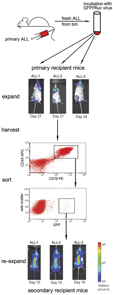
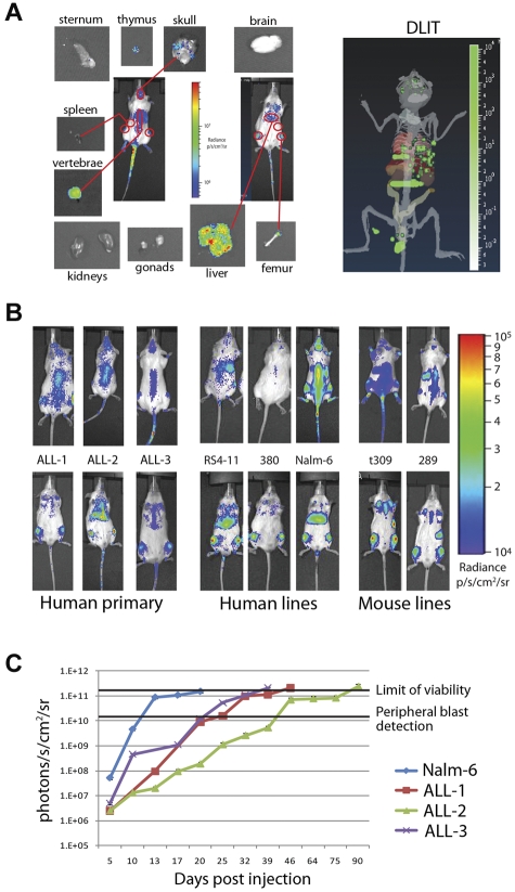
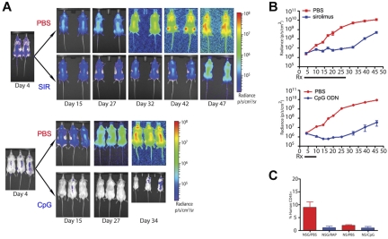
The efficient engraftment in immune-deficient mice achieved with both acute lymphoblastic leukemia (ALL) cell lines and primary samples has facilitated identification of the antileukemia activity of a wide variety of agents. Despite widespread usage, however, little is known about the early ALL localization and engraftment kinetics in this model, limiting experimental read-outs primarily to survival and endpoint analysis at high disease burden. In this study, we report that bioluminescent imaging can be reproducibly achieved with primary human ALL samples. This approach provides a noninvasive, longitudinal measure of leukemia burden and localization that enhances the sensitivity of treatment response detection and provides greater insight into the mechanism of action of antileukemia agents. In addition, this study reveals significant cell line- and species-related differences in leukemia migration, especially early in expansion, which may confound observations between various leukemia models. Overall, this study demonstrates that the use of bioluminescent primary ALL allows the detection and quantitation of treatment effects at earlier, previously unquantifiable disease burdens and thus provides the means to standardize and expedite the evaluation of anti-ALL activity in preclinical xenograft studies.


Figures

Figure 1
Figure 1
Our strategy to generate stable ffluc+ primary ALL samples. Bulk transduction of primary patient ALL, freshly obtained from NSG mouse bone marrow, via lentivirus results in low-level GFP/ffluc expression, which can be purified by flow sorting. Three primary patient ALL samples (ALL-1, ALL-2, and ALL-3) were harvested from moribund NSG mice, transduced overnight, and viable cells injected into primary recipients via tail vein. All 3 samples engrafted with a small GFP+ population, which was then purified using the gating strategy shown (sample ALL-1 sorting is shown). This enrichment results in stable, > 99% bioluminescent primary ALL with resultant early detection and evenly disseminated disease in secondary (shown in bottom panels) and later recipient mice.
Figure 2
Figure 2
Bioluminescence can be used to distinguish patterns and timing of organ involvement for both human and mouse leukemia. (A) Prone, supine, and organ images of an NSG mouse injected with 1 × 10⁶ ffluc+ primary ALL-1 cells. Outlined 2D regions of interest correspond to organ involvement validated at day 3 of engraftment by isolated imaging. DLIT image of mouse internal anatomy, with skin and gut removed for clarity, reconstructed from images is shown (right panel). Green voxels represent point sources of light within putative target organs, verified by ex vivo imaging and flow cytometry. (B) Day 3 intensity maps of NSG mice injected with 1 × 10⁶ ffluc+ leukemia cells as indicated. These representative mice show the differing patterns of organ involvement at this early time point. Human ALL cells establish within the liver and bone marrow (primary ALL, RS4-11, and Nalm-6) or bone marrow only (380), whereas both mouse cell lines studied rapidly home to the spleen and bone marrow (t309 and 289). (C) Time course of leukemia engraftment reveals consistent bioluminescent correlation with appearance of peripheral blasts and maximal moribund disease burden. Nalm-6 (20-22 days), primary ALL-1 (40-50 days), primary ALL-2 (80-100 days), and primary ALL-3 (35-40 days) all become moribund at 2 × 10¹¹ photons/s/cm2. Appearance of peripheral blasts (> 1%) happens consistently at systemic bioluminescent tumor burdens of > 1 × 10¹⁰ photons/s/cm2. In ALL-2, the stable bioluminescent burden from 46 to 75 days corresponds with stable but high peripheral blast burden (30%-50%) before a progression to end stage between 80 and 100 days. Results are presented as mean ± SE (n = 5 for primary ALL samples, n = 10 for Nalm-6).
Figure 3
Figure 3
Bioluminescence enables the visualization and quantification of therapeutic responses of primary ALL significantly earlier than other measurable parameters. (A) Intensity maps of total body disease developing over time in NSG mice treated with sirolimus (SIR) for 24 days (top panels) or in NOD/SCID mice treated with CpG ODN (CpG) over 8 days (bottom panels). Untreated (PBS) controls for both strains are shown. Results obtained with patient sample ALL-1 are shown. (B) Graphs with quantification of the bioluminescent ALL responses over time shown in panel A to treatment with sirolimus (top panel) or CpG ODN (bottom panel). (C) Flow cytometric evaluation of peripheral blood ALL burden at day 45 in the control, sirolimus-treated, and CpG ODN-treated mice shown in panels A and B. Overall, this experiment is not designed to compare the treatment strategies but rather to demonstrate the additional detail of ALL treatment response revealed by bioluminescent imaging.

References

    1. Bernardi R, Grisendi S, Pandolfi PP. Modelling haematopoietic malignancies in the mouse and therapeutical implications. Oncogene. 2002;21(21):3445–3458. - PubMed
    1. Baersch G, Mollers T, Hotte A, et al. Good engraftment of B-cell precursor ALL in NOD-SCID mice. Klin Padiatr. 1997;209(4):178–185. - PubMed
    1. Lapidot T, Fajerman Y, Kollet O. Immune-deficient SCID and NOD/SCID mice models as functional assays for studying normal and malignant human hematopoiesis. J Mol Med. 1997;75(9):664–673. - PubMed
    1. Ito M, Hiramatsu H, Kobayashi K, et al. NOD/SCID/gamma(c)(null) mouse: an excellent recipient mouse model for engraftment of human cells. Blood. 2002;100(9):3175–3182. - PubMed
    1. Liem NL, Papa RA, Milross CG, et al. Characterization of childhood acute lymphoblastic leukemia xenograft models for the preclinical evaluation of new therapies. Blood. 2004;103(10):3905–3914. - PubMed

Publication types

MeSH terms

Substances