

Reduced NO signaling during pregnancy attenuates outward uterine artery remodeling by altering MMP expression and collagen and elastin deposition
- PMID: 21856919
- PMCID: PMC3197352
- DOI: 10.1152/ajpheart.00519.2011
Reduced NO signaling during pregnancy attenuates outward uterine artery remodeling by altering MMP expression and collagen and elastin deposition
Abstract
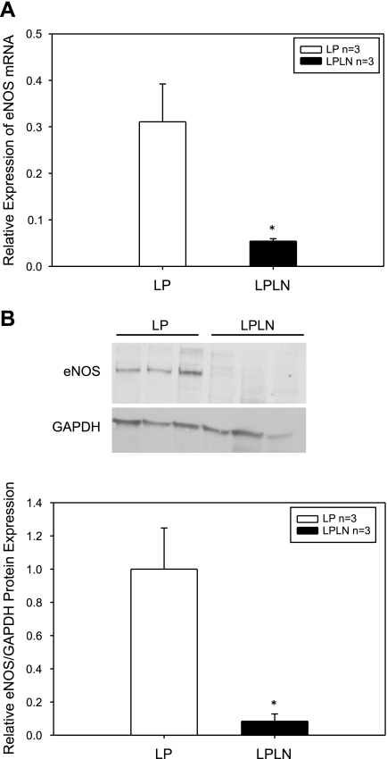
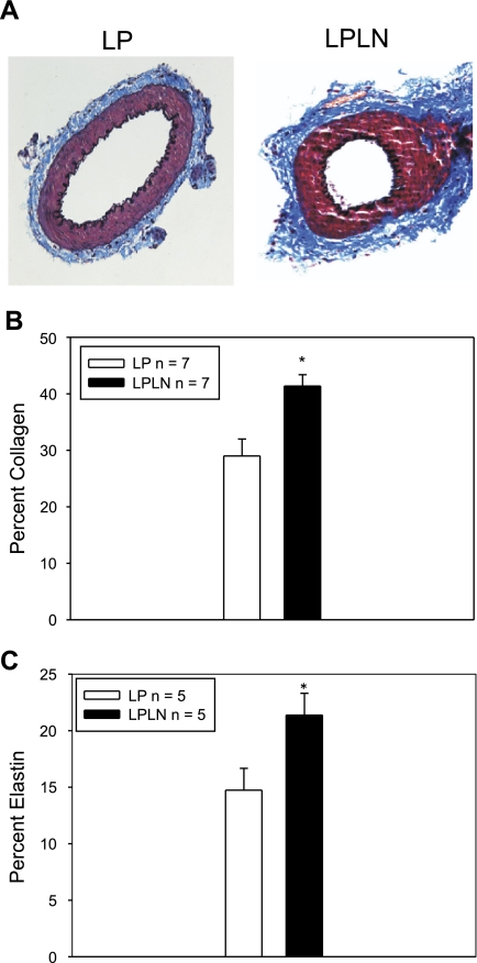
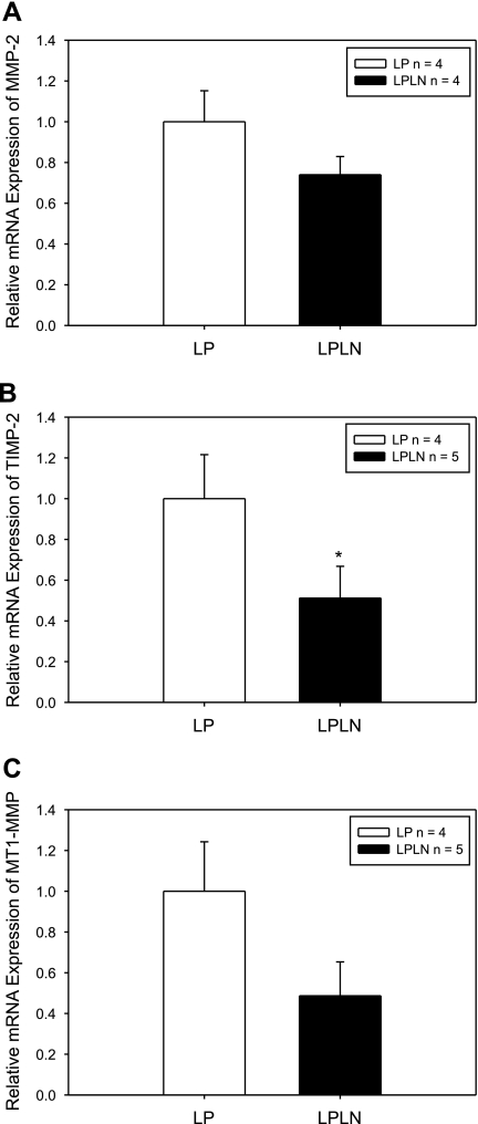
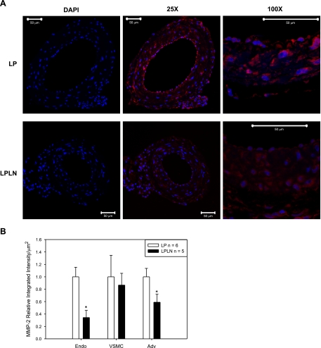
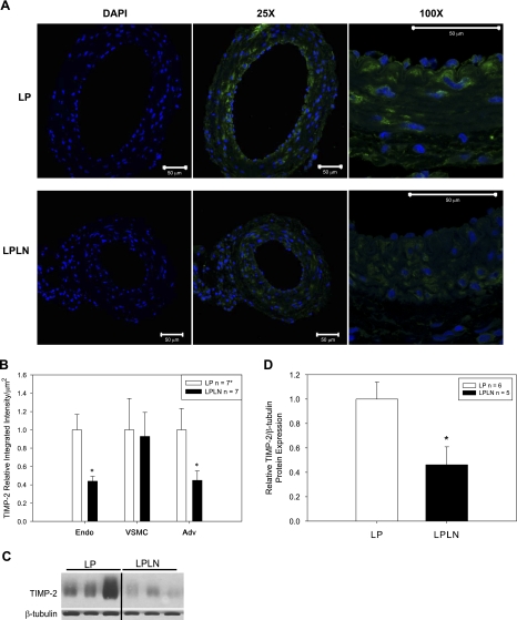
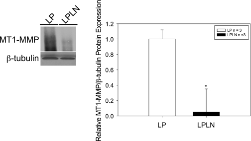
Recent findings indicate that endothelial nitric oxide (NO) plays a key role in uterine artery outward circumferential remodeling during pregnancy. Although the underlying mechanisms are not known, they likely involve matrix metalloproteinases (MMPs). The goal of this study was to examine the linkage among NO inhibition, expansive remodeling, and MMP expression within the uterine vascular wall. Adult female rats were treated with N(G)-nitro-L-arginine methyl ester [L-NAME (LPLN)] beginning on day 10 of pregnancy and until death at day 20 and compared with age-matched controls [late pregnant (LP)]. Mean arterial pressure of LPLN rats was significantly higher than controls. LPLN fetal and placental weights were significantly reduced compared with controls. Main uterine arteries (mUA) were collected to determine dimensional properties (lumen area and wall thickness), collagen and elastin content, and levels of endothelial nitric oxide synthase (eNOS) and MMP expression. Circumferential remodeling was attenuated, as evidenced by significantly smaller lumen diameters. eNOS RNA and protein were significantly (>90%) decreased in the LPLN mUA compared with LP. Collagen and elastin contents were significantly increased in LPLN rats by ∼10 and 25%, respectively, compared with LP (P < 0.05). Both MMP-2 and tissue inhibitors of metalloproteinase-2 as assessed by immunofluorescence were lower in the endothelium (reduction of 60%) and adventitia (reduction of 50%) of LPLN compared with LP mUA. Membrane bound MMP-1 (MT1-MMP) as assessed by immunoblot was significantly decreased in LPLN. These data suggest a novel contribution of MMPs to gestational uterine vascular remodeling and substantiate the linkage between NO signaling and gestational remodeling of the uterine circulation via altered MMP, TIMP-2, and MT1-MMP expression and activity.
Figures
Similar articles
-
Buckling Reduces eNOS Production and Stimulates Extracellular Matrix Remodeling in Arteries in Organ Culture.Ann Biomed Eng. 2016 Sep;44(9):2840-50. doi: 10.1007/s10439-016-1571-0. Epub 2016 Feb 25. Ann Biomed Eng. 2016. PMID: 26913855 Free PMC article.
-
Decreased uterine vascularization and uterine arterial expansive remodeling with reduced matrix metalloproteinase-2 and -9 in hypertensive pregnancy.Am J Physiol Heart Circ Physiol. 2020 Jan 1;318(1):H165-H180. doi: 10.1152/ajpheart.00602.2019. Epub 2019 Dec 13. Am J Physiol Heart Circ Physiol. 2020. PMID: 31834839 Free PMC article.
-
Coordinated elevation of membrane type 1-matrix metalloproteinase and matrix metalloproteinase-2 expression in rat uterus during postpartum involution.Reprod Biol Endocrinol. 2006 Jun 2;4:32. doi: 10.1186/1477-7827-4-32. Reprod Biol Endocrinol. 2006. PMID: 16740171 Free PMC article.
-
Matrix metalloproteinases as drug targets in preeclampsia.Curr Drug Targets. 2013 Mar;14(3):325-34. doi: 10.2174/1389450111314030004. Curr Drug Targets. 2013. PMID: 23316964 Free PMC article. Review.
-
Physiological remodelling of the maternal uterine circulation during pregnancy.Basic Clin Pharmacol Toxicol. 2012 Jan;110(1):12-8. doi: 10.1111/j.1742-7843.2011.00793.x. Epub 2011 Oct 21. Basic Clin Pharmacol Toxicol. 2012. PMID: 21902814 Review.
Cited by
-
Buckling Reduces eNOS Production and Stimulates Extracellular Matrix Remodeling in Arteries in Organ Culture.Ann Biomed Eng. 2016 Sep;44(9):2840-50. doi: 10.1007/s10439-016-1571-0. Epub 2016 Feb 25. Ann Biomed Eng. 2016. PMID: 26913855 Free PMC article.
-
Potassium Channels in the Uterine Vasculature: Role in Healthy and Complicated Pregnancies.Int J Mol Sci. 2022 Aug 21;23(16):9446. doi: 10.3390/ijms23169446. Int J Mol Sci. 2022. PMID: 36012712 Free PMC article. Review.
-
Coronary microvascular disease as an early culprit in the pathophysiology of diabetes and metabolic syndrome.Pharmacol Res. 2017 Sep;123:114-121. doi: 10.1016/j.phrs.2017.07.004. Epub 2017 Jul 9. Pharmacol Res. 2017. PMID: 28700893 Free PMC article. Review.
-
Association of parity with carotid diameter and distensibility: multi-ethnic study of atherosclerosis.Hypertension. 2014 Aug;64(2):253-8. doi: 10.1161/HYPERTENSIONAHA.114.03285. Hypertension. 2014. PMID: 24842921 Free PMC article.
-
The role of reactive oxygen species in microvascular remodeling.Int J Mol Sci. 2014 Dec 19;15(12):23792-835. doi: 10.3390/ijms151223792. Int J Mol Sci. 2014. PMID: 25535075 Free PMC article. Review.
References
-
- Arribas SM, Hinek A, Gonzalez MC. Elastic fibres and vascular structure in hypertension. Pharmacol Ther 111: 771–791, 2006 - PubMed
-
- Bankowski E, Romanowicz L, Jaworski S. Collagen of umbilical cord arteries and its alterations in EPH-gestosis. J Perinat Med 21: 491–498, 1993 - PubMed
-
- Buhimschi I, Yallampalli C, Chwalisz K, Garfield RE. Pre-eclampsia-like conditions produced by nitric oxide inhibition: effects of l-arginine, d-arginine and steroid hormones. Hum Reprod 10: 2723–2730, 1995 - PubMed
-
- Buhimschi IA, Saade GR, Chwalisz K, Garfield RE. The nitric oxide pathway in pre-eclampsia: pathophysiological implications. Hum Reprod Update 4: 25–42, 1998 - PubMed
-
- Chakraborti S, Mandal M, Das S, Mandal A, Chakraborti T. Regulation of matrix metalloproteinases: an overview. Mol Cell Biochem 253: 269–285, 2003 - PubMed
Publication types
MeSH terms
Substances
Grants and funding
LinkOut - more resources
Full Text Sources
Medical
Research Materials
Miscellaneous