. 2011 Nov;301(5):H1907-15.
doi: 10.1152/ajpheart.00397.2011. Epub 2011 Aug 19.

Alignment of sarcoplasmic reticulum-mitochondrial junctions with mitochondrial contact points

Affiliations

Alignment of sarcoplasmic reticulum-mitochondrial junctions with mitochondrial contact points

Cecília García-Pérez et al. Am J Physiol Heart Circ Physiol. 2011 Nov.

Abstract

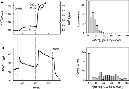
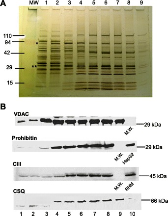
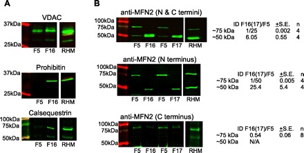
Propagation of ryanodine receptor (RyR2)-derived Ca(2+) signals to the mitochondrial matrix supports oxidative ATP production or facilitates mitochondrial apoptosis in cardiac muscle. Ca(2+) transfer likely occurs locally at focal associations of the sarcoplasmic reticulum (SR) and mitochondria, which are secured by tethers. The outer mitochondrial membrane and inner mitochondrial membrane (OMM and IMM, respectively) also form tight focal contacts (contact points) that are enriched in voltage-dependent anion channels, the gates of OMM for Ca(2+). Contact points could offer the shortest Ca(2+) transfer route to the matrix; however, their alignment with the SR-OMM associations remains unclear. Here, in rat heart we have studied the distribution of mitochondria-associated SR in submitochondrial membrane fractions and evaluated the colocalization of SR-OMM associations with contact points using transmission electron microscopy. In a sucrose gradient designed for OMM purification, biochemical assays revealed lighter fractions enriched in OMM only and heavier fractions containing OMM, IMM, and SR markers. Pure OMM fractions were enriched in mitofusin 2, an ~80 kDa mitochondrial fusion protein and SR-mitochondrial tether candidate, whereas in fractions of OMM + IMM + SR, a lighter (~50 kDa) band detected by antibodies raised against the NH(2) terminus of mitofusin 2 was dominating. Transmission electron microscopy revealed mandatory presence of contact points at the junctional SR-mitochondrial interface versus a random presence along matching SR-free OMM segments. For each SR-mitochondrial junction at least one tether was attached to contact points. These data establish the contact points as anchorage sites for the SR-mitochondrial physical coupling. Close coupling of the SR, OMM, and IMM is likely to provide a favorable spatial arrangement for local ryanodine receptor-mitochondrial Ca(2+) signaling.


Figures

Fig. 1.
Fig. 1.
Sarcoplasmic reticulum (SR)-derived Ca2+ signal reception and matrix dehydrogenase activation are preserved in isolated rat heart mitochondria (mito). A: [Ca2+]m and [Ca2+]c (measured with rhod2 accumulated to the mitochondrial matrix and fura 2 added to the cytosolic buffer, respectively) responses were simultaneously imaged in the mitochondrial fractions (10,000 g pellet) of rat heart homogenates. The stimulation protocol was sequential addition of caffeine [Caf; 10 mM, that evokes maximum ryanodine receptor (RyR2)-mediated Ca2+ release from the SR] and a reference Ca2+ pulse (20 μM CaCl2) that elevated [Ca2+]c to ∼7 μM. To maximize RyR2-mediated Ca2+ mobilization from the SR, caffeine was applied together with the sarco(endo)plasmic reticulum Ca2+-ATPase (SERCA) inhibitor thapsigargin (Tg; 10 μM). B: in parallel experiments, the changes in NAD(P)H autofluorescence were recorded. The traces of [Ca2+]m and NAD(P)H are means of ∼100 mitochondria. The histograms at right show the distribution of responses to caffeine stimulation normalized to the reference Ca2+ pulse amongst 100 randomly selected mitochondrial spots on the field. Mitochondria not showing response to either caffeine or the Ca2+ reference pulse were excluded from the analysis.
Fig. 2.
Fig. 2.
Purification of outer mitochondrial membrane (OMM) and OMM-inner mitochondrial membrane (IMM) contacts from rat heart mitochondria. Mitochondria isolated from rat heart homogenates were subfractionated on a discontinuous sucrose gradient as the scheme shows at left (with sucrose concentrations shown as percent weight per volume). Protein concentration profile (bottom) and normalized (to the maximum, max) activities (top) of monoaminooxidase (MAO), an OMM resident, and succinate dehydrogenase (SDH), an IMM localized enzyme in the isolated subfractions, are shown. Data are means ± range from 2 independent preparations (each from 2 hearts).
Fig. 3.
Fig. 3.
SDS-PAGE and Western blot analysis of proteins in the mitochondrial subfractions. A: silver nitrate-stained protein bands after running the mitochondrial subfractions (1–9) on a 4–15% gradient gel. The lane labeled MW was loaded with molecular weight markers. *Predicted bands for hexokinase (HK); **predicted bands for voltage-dependent anion channel (VDAC) with the adenine nucleotide translocator of IMM (ANT). B: representative Western blots of VDAC (OMM marker), prohibitin (IMM marker), respiratory complex III (CIII; IMM marker), and calsequestrin (CSQ; SR marker). RHM, rat heart mitochondrial fraction.
Fig. 4.
Fig. 4.
Purification of OMM-associated SR from rat heart mitochondria. Submitochondrial fractions (20–24) were separated on a sucrose density gradient. The approximate mapping of the fractions to the sucrose density levels is illustrated by the scheme at left. A: bimodal protein profile is shown at the bottom; enzyme activities of HK (○; OMM marker), MAO (●; OMM marker), and SDH (squares; IMM marker) in the subfractions (normalized to maximum) are shown at top. Data are means ± SE from 3 preparations (HK, SDH) or means ± range from 2 preparations (MAO). B: representative Western blots of CIII (IMM marker) and SERCA2a from the submitochondrial fractions shown in A.
Fig. 5.
Fig. 5.
Correlating junctional SR(jSR)-mitochondria junctions and SR-mitochondrial tethers with OMM-IMM contact points using transmission electron microscopy (TEM). Cardiac muscle samples from rat left ventricle were fixed and processed for TEM. High-magnification (76,000× at 7 inches print size) images were taken from longitudinal as well as transversal sections of regions containing well recognizable dyads and mitochondria. A: representative high-magnification (left) TEM image of a jSR-OMM junction showing the cross section of a T-tubule (T) and a jSR sack (SR) in association with a mitochondrion (M). Note the contact points (cp) between the OMM and IMM and the discrete electron densities interconnecting the SR and OMM corresponding to tethers (white arrows: the bottom one attaching to a cp, whereas the top one attaches to an OMM segment free of IMM). The high-magnification image at left is zoomed out by a factor of 10× at right to show the organellar context. B: bar graph analyzing the likelihood of an OMM-IMM contact point being located at a jSR-OMM interface or at size-matched SR-free OMM segments selected as shown in the scheme. For each segment the presence and absence of a contact point scores 1 and 0, respectively. For each mitochondrion there is one OMM-jSR junction, and 1–3 SR-free OMM segments are averaged (in some cases it was not possible to have 3 SR-free OMM segments equal in size to the jSR-OMM junction and having well recognizable membranes). Nineteen mitochondria from 3 animals were analyzed. The difference between the 2 groups is significant (P < 0.005, using Wilcoxon signed rank test). C: bar graph summarizing the number of SR-OMM tethers observed in each jSR-mitochondria junction that ended at an OMM-IMM contact point or at an IMM-free OMM region. Data are means ± SE (n = 19; P > 0.05 using Wilcoxon signed rank test).
Fig. 6.
Fig. 6.
Determination of mitofusin 2 (MFN2) protein in the light and heavy submitochondrial fractions. Light and heavy fractions close to the 2 protein peaks (fractions 5 and 16–17, respectively, from gradients separated to 22 to 23 fractions in total) were compared in their MFN2 immunoreactivity via Western blot. Reactions were visualized using the infrared fluorescence LI-COR detection system. A: VDAC (∼31 kDa), prohibitin (∼29 kDa), and calsequestrin (∼60 kDa) antibodies were used to confirm the membrane composition of the fractions (note the omnipresence of VDAC and the enrichment of prohibitin and calsequestrin bands in fraction 16). The crude RHM was used for reference (positive control). B: MFN2 (∼80 kDa) immunoreactive bands detected by 3 different antibodies recognizing either the COOH terminus (bottom), NH2 terminus (middle), or both (top) in the subfractions shown in A and in fractions 5 and 17 (F5, F17) from another preparation. Note the lack or decreased presence of the ∼75 kDa band in the heavy fractions and at the same time the appearance of a lower MW band (∼50 kDa). The double-banding in F16/17 when using antibody against the N and C termini of MFN2 (top) is most likely due to a cross-reaction, and it is not an artifact since it also appears in mitochondrial preparations of rat skeletal muscle (not shown). The numbers on the right show the integrated fluorescence intensity [integrated density (ID)] of F16 relative to F5 for the ∼75 kDa and ∼50 kDa bands.
Fig. 7.
Fig. 7.
Functional consequences of the alignment of jSR-OMM junctions with mitochondrial contact points. The scheme illustrates the potential advantages of aligning the OMM-anchored jSR with mitochondrial contact points with regard to the efficacy of local Ca2+ transfer to the mitochondrial matrix. The top depicts a nonaligned scenario, whereas the bottom shows the aligned arrangement. MCU, mitochondrial Ca2+ uniporter; PT, permeability transition.

References

    1. Adams V, Bosch W, Schlegel J, Wallimann T, Brdiczka D. Further characterization of contact sites from mitochondria of different tissues: topology of peripheral kinases. Biochim Biophys Acta 981: 213–225, 1989 - PubMed
    1. Ardail D, Gasnier F, Lerme F, Simonot C, Louisot P, Gateau-Roesch O. Involvement of mitochondrial contact sites in the subcellular compartmentalization of phospholipid biosynthetic enzymes. J Biol Chem 268: 25985–25992, 1993 - PubMed
    1. Ardail D, Privat JP, Egret-Charlier M, Levrat C, Lerme F, Louisot P. Mitochondrial contact sites. Lipid composition and dynamics. J Biol Chem 265: 18797–18802, 1990 - PubMed
    1. Balaban RS. Domestication of the cardiac mitochondrion for energy conversion. J Mol Cell Cardiol 46: 832–841, 2009 - PMC - PubMed
    1. Bathori G, Csordas G, Garcia-Perez C, Davies E, Hajnoczky G. Ca2+-dependent control of the permeability properties of the mitochondrial outer membrane and voltage-dependent anion-selective channel (VDAC). J Biol Chem 281: 17347–17358, 2006 - PubMed

Publication types

MeSH terms