. 2011 Sep 15;187(6):3177-85.
doi: 10.4049/jimmunol.1100206. Epub 2011 Aug 19.

G alpha i2 and ZAP-70 mediate RasGRP1 membrane localization and activation of SDF-1-induced T cell functions

Affiliations

G alpha i2 and ZAP-70 mediate RasGRP1 membrane localization and activation of SDF-1-induced T cell functions

Kimberly N Kremer et al. J Immunol. .

Abstract

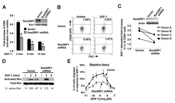
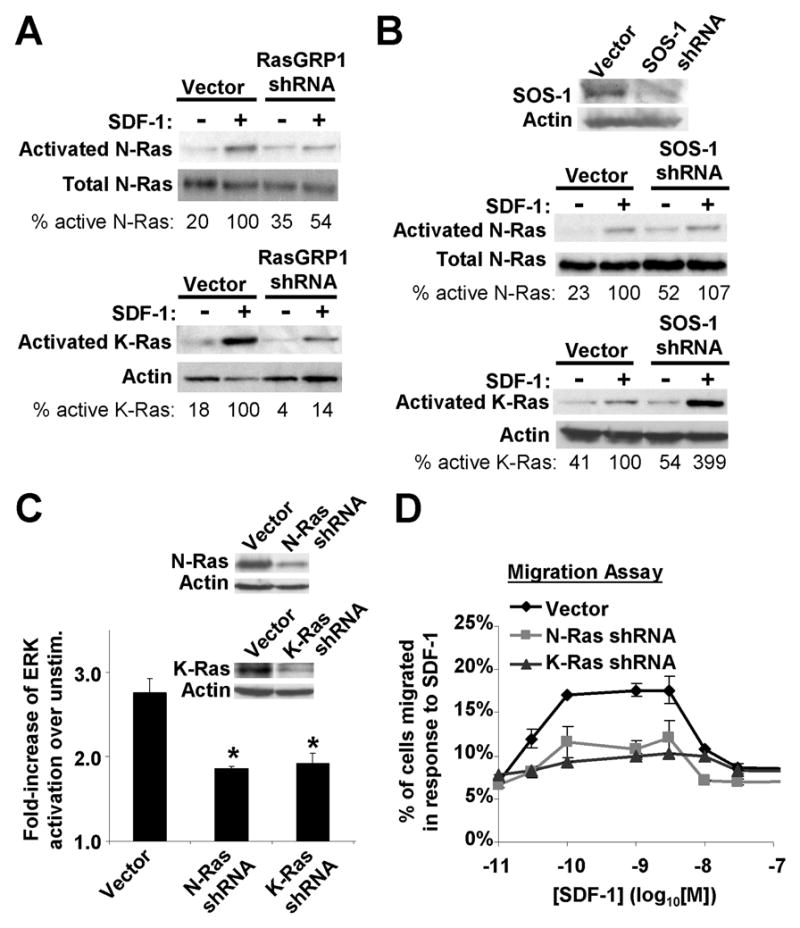
RasGRP1, a Ras guanine-nucleotide exchange factor, critically mediates T cell development and function and controls immunodeficiency and autoimmunity. In this study, we describe a unique mechanism of mobilization and activation of RasGRP1 in response to SDF-1, a chemokine that signals via the G protein-coupled receptor CXCR4. Depletion of RasGRP1 impaired SDF-1-stimulated human T cell migration, expression of the activation marker CD69, and activation of the ERK MAPK pathway, indicating that RasGRP1 mediates SDF-1 functions. SDF-1 treatment caused RasGRP1 to localize to the plasma membrane to activate K-Ras and to the Golgi to activate N-Ras. These events were required for cellular migration and for ERK activation that mediates downstream transcriptional events in response to SDF-1. SDF-1-dependent localization of RasGRP1 did not require its diacylglycerol-binding domain, even though diacyglycerol was previously shown to mediate localization of RasGRP1 in response to Ag stimulation. This domain was, however, required for activity of RasGRP1 after its localization. Intriguingly, SDF-1 treatment of T cells induced the formation of a novel molecular signaling complex containing RasGRP1, Gαi2, and ZAP-70. Moreover, SDF-1-mediated signaling by both Gi proteins and ZAP-70 was required for RasGRP1 mobilization. In addition, RasGRP1 mobilization and activation in response to SDF-1 was dependent on TCR expression, suggesting that CXCR4 heterodimerizes with the TCR to couple to ZAP-70 and mobilize RasGRP1. These results increase understanding of the molecular mechanisms that mediate SDF-1 effects on T cells and reveal a novel mechanism of RasGRP1 regulation. Other G protein-coupled receptors may similarly contribute to regulation of RasGRP1.


Figures

Fig. 1
Fig. 1. RasGRP1 is required for SDF-1/CXCL12 signaling to stimulate the ERK MAP kinase pathway, CD69 expression, and migration of T cells
A) Jurkat T cells transfected with either a RasGRP1 shRNA or vector alone were stimulated with SDF-1 and assayed for ERK activation. Bars denote the fold-increase in ERK activation of SDF-1-stimulated as compared to unstimulated cells ± S.E.M., n = 6; *, significantly different from control, p<0.05. Inset: Immunoblot of cell lysates showing decreased RasGRP1 compared to a control protein (Vav-1). B) Normal, primary human T cells (PBMC T cells) transfected with RasGRP1 shRNA or vector alone were stimulated for 24 hr with SDF-1 and assayed for CD69 expression by flow cytometry. C) Summary of multiple experiments performed as in B), using T cells from 4 donors. Inset: Immunoblot of cell lysates. D&E) Jurkat cells were transfected with RasGRP1 shRNA as in A), and assayed for active GTP bound Rho or total Rho (D), or migration (E) in response to SDF-1. The relative % of Rho activation (D) was determined by normalizing to total Rho. Each point in E) denotes the mean % of cells migrated ± S.D., n=3. Results shown are representative of 3 independent experiments.
Fig. 2
Fig. 2. SDF-1/CXCR4 signaling utilizes the TCR to mobilize RasGRP1 in order to enhance activation of N- and K- Ras
A–D) Live, individual PBMC T cells or Jurkat cells expressing a fluorescent fusion protein of RasGRP1 (RasGRP1-YFP) were analyzed by confocal microscopy ± SDF-1. Arrows indicate RasGRP1-YFP localized to the Golgi and plasma membranes in different z-slices. Bars here & below, 2 μm. A differential interference contrast (DIC) image is included in B). Where indicated, cells also express the Golgi marker, GalT-CFP (blue). A) n=29 PBMC T cells, B) n=7 PBMC T cells, C) n=4 Jurkat cells, D), n=49 Jurkat cells. E) Cell-surface expression of the TCR on TCR-βdeficient & TCR-β-reconstituted Jurkat cells. F) TCR-β-deficient cells assayed for RasGRP1 localization as in Fig. 2D. G) Summary of multiple cells assayed as in D&F), n=24–49 for each bar. H–I) Jurkat, TCR-β-deficient, and TCR-β-reconstituted cells were assayed for active, GTP-bound Ras isoforms in response to SDF-1. For controls, cell lysates were immunoblotted for total N-Ras or actin. The % of the indicated active Ras isoform was determined (here and below) by normalizing to the indicated loading control. J) Jurkat or TCR-β-deficient cells expressing a GTP-Ras-binding domain fused to YFP (RBD-YFP) and either N-Ras, K-RasB, or H-Ras were imaged as live cells by confocal microscopy ± SDF-1. K) Results of analyzing multiple cells as in J: n=15–20 per bar.
Fig. 3
Fig. 3. N-Ras and K-Ras mediate ERK activation and migration of T cells via a mechanism requiring RasGRP1 but not SOS-1
A) RasGRP1 was depleted from Jurkat cells as in Fig. 1A and N- or K-Ras activation in response to 2 min SDF-1 was assayed as in Fig. 2H, n = 3. B) Lower gels, SOS-1 was depleted from Jurkat cells via shRNA and N- and K-Ras activation was assayed as in A). Upper gel, cell lysates immunoblotted for SOS-1. n=3. C) N- or K-Ras were specifically depleted from Jurkat cells using shRNA, and ERK activation in response to 8 min of SDF-1 was assayed as in Fig. 1A. Bars denote the fold increase in ERK activation of stimulated as compared to unstimulated cells ± S.E.M., n=5; *, significantly different from controls, p<0.05. Inset: Immunoblot of N- and K-Ras compared to actin (control) in cell lysates. D) N- or K-Ras were depleted from Jurkat cells as in C), and migration was assayed as in Fig. 1E. The experiment shown is representative of 3 independent experiments.
Fig. 4
Fig. 4. The diacylglycerol binding domain of RasGRP1 is not required for its membrane localization in response to SDF-1, but is required for its activity
A) The localization of RasGRP1-ΔDAG-YFP expressed in PBMC T cells was assayed as in Fig. 2A; n = 31–33. B) Jurkat cells were transfected with the indicated vector, 72 hr later the cells were lysed and RasGRP1 was immunoprecipitated and immunoblotted. Whole cell lysates were immunblotted with actin (control). C) Jurkat cells expressing RasGRP1 shRNA alone, or together with shRNA-resistant RasGRP1 (either RasGRP1WT or RasGRP1-ΔDAG), were stimulated for 8 min with SDF-1 and assayed for ERK as in Fig 1A. n=3; * or **, significantly different, p<0.05. GFP gating for this experiment is shown in Supplemental Figure 3.
Fig. 5
Fig. 5. Gαi2 and ZAP-70 mediate the localization of RasGRP1 and thus lead to the activation of N-Ras and K-Ras
A&B) Jurkat cells were stimulated with SDF-1, lysed, and either RasGRP1, Gαi2, or ZAP-70 were immunoprecipitated and immunoblotted to reveal co-purifying proteins, n=3. C) PBMC T cells expressing RasGRP1WT-YFP were pretreated with either the control PTX-B toxin or PTX and assayed for RasGRP1 localization as in Fig. 2A. D) Summary of multiple cells assayed as in C), n=33–38 for each bar. E) Jurkat cells were pretreated with either PTX-B or PTX and assayed for N- or K-Ras activation in response to 2 min SDF-1 as in Fig. 2H, n=3. F) Summary of multiple PBMC T cells expressing RasGRP1WT-YFP, that were pretreated with vehicle (DMSO) or piceatannol and assayed for RasGRP1 localization as in Fig. 2A, n=25–32 for each bar. G) ZAP-70-deficient Jurkat cells were assayed for RasGRP1 localization as in Fig. 2D, n=12. H) Normal Jurkat, ZAP-70-deficient, or ZAP-70-reconstituted Jurkat cells were assayed for N- or K-Ras in response to 2 min SDF-1 as in Fig. 2H, n=3.
Fig. 6
Fig. 6. SDF-1 utilizes a novel mechanism to regulate RasGRP1 localization and activation in order to modulate T cell functions
Based on our results, we propose the model shown in Fig. 6. SDF-1 binding to CXCR4 induces formation of the CXCR4-TCR heterodimeric receptor which signals to cause the binding of Gαi2 and ZAP-70 to RasGRP1. This RasGRP1-Gαi2-ZAP-70 complex allows for mobilization of RasGRP1 to the plasma membrane and the Golgi. Following localization of RasGRP1, DAG binds to the DAG-binding domain of RasGRP1 thereby activating RasGRP1, which in turn activates K-Ras at the plasma membrane and N-Ras at the Golgi. Active K-Ras and N-Ras then lead to ERK activation and subsequent gene transcription as well as Rho activation that leads to T cell migration.

Similar articles

Cited by

References

    1. Trampont PC, Tosello-Trampont AC, Shen Y, Duley AK, Sutherland AE, Bender TP, Littman DR, Ravichandran KS. CXCR4 acts as a costimulator during thymic beta-selection. Nature immunology. 2010;11:162–170. - PMC - PubMed
    1. Lusso P. HIV and the chemokine system: 10 years later. The EMBO journal. 2006;25:447–456. - PMC - PubMed
    1. Suzuki Y, Rahman M, Mitsuya H. Diverse transcriptional response of CD4(+) T cells to stromal cell-derived factor (SDF)-1: cell survival promotion and priming effects of SDF-1 on CD4(+) T cells. J Immunol. 2001;167:3064–3073. - PubMed
    1. Kumar A, Humphreys TD, Kremer KN, Bramati PS, Bradfield L, Edgar CE, Hedin KE. CXCR4 physically associates with the T cell receptor to signal in T cells. Immunity. 2006;25:213–224. - PubMed
    1. Kremer KN, Kumar A, Hedin KE. Haplotype-independent costimulation of IL-10 secretion by SDF-1/CXCL12 proceeds via AP-1 binding to the human IL-10 promoter. J Immunol. 2007;178:1581–1588. - PMC - PubMed

Publication types

MeSH terms

Substances