

Alterations in T2 relaxation magnetic resonance imaging of the ovine intervertebral disc due to nonenzymatic glycation
- PMID: 21857410
- PMCID: PMC3348580
- DOI: 10.1097/BRS.0b013e31822ce81f
Alterations in T2 relaxation magnetic resonance imaging of the ovine intervertebral disc due to nonenzymatic glycation
Abstract
Study design: An in vitro study using ovine intervertebral discs to correlate the effects of increasing advanced glycation end-products (AGEs) with disc hydration evaluated by magnetic resonance imaging (MRI).
Objective: To determine the relationship between the level of AGEs and tissue water content in intervertebral discs using T2 relaxation MRI.
Summary of background data: AGEs result from nonenzymatic glycation, and AGEs have been shown to accumulate in the intervertebral disc tissue with aging and degeneration. AGEs can alter biochemical properties, including the hydrophobicity of the extracellular matrix. Because one of the degenerative signs of the intervertebral disc (IVD) is reduced hydration, we hypothesized that increased levels of tissue AGEs contribute to disc hydration. T2 relaxation MRI has been shown to be sensitive to the hydration status of the disc and may be valuable in detecting the changes in the IVD mediated by the increase of AGEs.
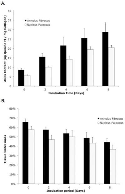
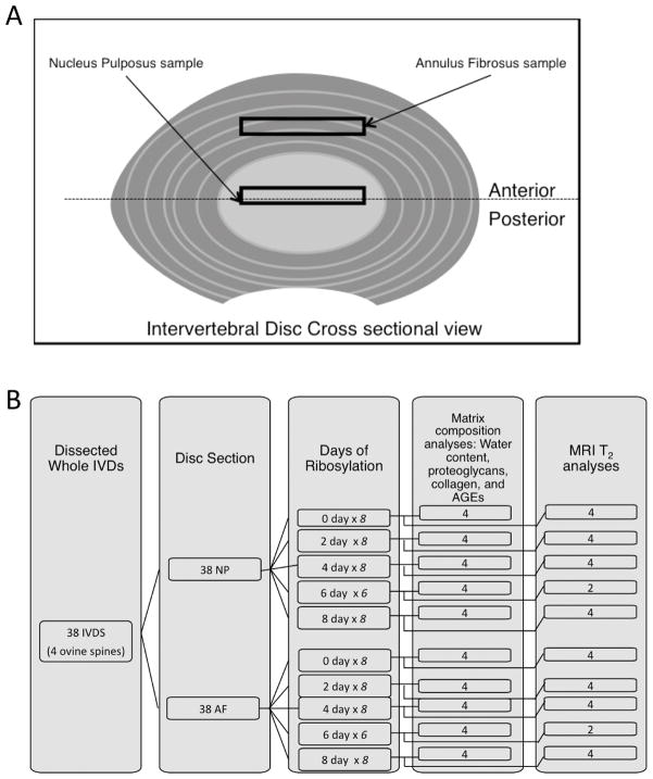
Methods: Thirty-eight IVDs were obtained from 4 ovine spines, and the annulus fibrosis (AF) and nucleus pulposus (NP) tissues were isolated from these discs. The tissues were incubated in either a ribosylation or control solution for up to 8 days to induce the formation of AGEs. T2 relaxation times were obtained from these tissues after ribosylation. These tissues were subsequently analyzed for hydration, proteoglycan, collagen, and AGEs content.
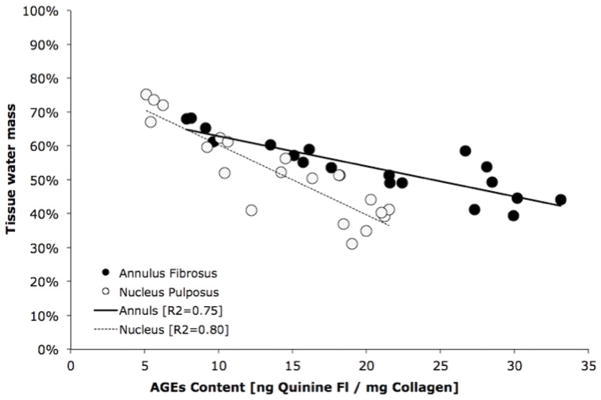
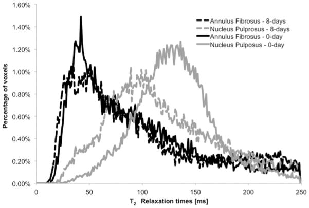
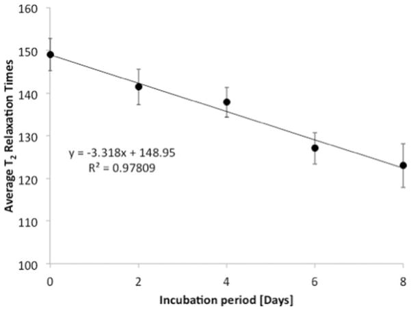
Results: In vitro ribosylation led to the increased accumulation of AGEs and reduced water content in both the AF and NP in a dose-dependent manner, but did not affect the proteoglycan and collagen composition. When analyzed by MRI, ribosylation significantly altered the mean T2 relaxation times in the NP (P = 0.001), but not in the AF (P = 0.912). Furthermore, the mean T2 values in the NP significantly decreased with increasing periods of incubation time (P < 0.001).
Conclusion: This study demonstrates that levels of AGEs in the IVD may affect the tissue water content. Moreover, these ribosylation-mediated changes in tissue hydration were detectable using T2 relaxation MRI. T2 relaxation MRI may provide a noninvasive tool to measure in vivo changes in disc hydration that are negatively correlated with the accumulation of AGEs.
Figures
References
-
- United States Bone and Joint Decade: The Burden of Musckoskeletal Diseases in the United States. Amer Acad Ortho Surg. 2008;Ch 9
-
- Praemer A, Furner S, Rice DP. Costs of musculoskeletal conditions in the US. Amer Acad Ortho Surg. 1992:145–170.
-
- Yokosuka K, Park JS, Jimbo K, et al. Advanced glycation end-products downregulating intervertebral disc cell production of proteoglycans in vitro. J Neurosurg: Spine. 2006;5(4):324–9. - PubMed
-
- Reiser KM. Nonenzymatic glycation of collagen in aging and diabetes. Proc Soc Exp Biol Med. 1991;196(1):17–29. - PubMed
Publication types
MeSH terms
Substances
Grants and funding
LinkOut - more resources
Full Text Sources
Medical
Research Materials
Miscellaneous
